Credit: Panagiotis Kastritis
Biochemists at Martin Luther University Halle-Wittenberg (MLU) have used a standard electron cryo-microscope to achieve surprisingly good images that are on par with those taken by far more sophisticated equipment. They have succeeded in determining the structure of ferritin almost at the atomic level. Their results were published in the journal PLOS ONE.
Electron cryo-microscopy has become increasingly important in recent years, especially in shedding light on protein structures. The developers of the new technology were awarded the Nobel Prize for Chemistry in 2017. The trick: the samples are flash frozen and then bombarded with electrons. In the case of traditional electron microscopy, all of the water is first extracted from the sample. This is necessary because the investigation takes place in a vacuum, which means water would evaporate immediately and make imaging impossible. However, because water molecules play such an important role in biomolecules, especially in proteins, they cannot be examined using traditional electron microscopy. Proteins are among the most important building blocks of cells and perform a variety of tasks. In-depth knowledge of their structure is necessary in order to understand how they work.
The research group led by Dr Panagiotis Kastritis, who is a group leader at the Centre for Innovation Competence HALOmem and a junior professor at the Institute of Biochemistry and Biotechnology at MLU, acquired a state-of-the-art electron cryo-microscope in 2019. “There is no other microscope like it in Halle,” says Kastritis. The new “Thermo Fisher Glacios 200 kV”, financed by the Federal Ministry of Education and Research, is not the best and most expensive microscope of its kind. Nevertheless, Kastritis and his colleagues succeeded in determining the structure of the iron storage protein apoferritin down to 2.7 ångströms (Å), in other words, almost down to the individual atom. One ångström equals one-tenth of a nanometre. This puts the research group in a similar league to departments with far more expensive equipment. Apoferritin is often used as a reference protein to determine the performance levels of corresponding microscopes. Just recently, two research groups broke a new record with a resolution of about 1.2 Å. “Such values can only be achieved using very powerful instruments, which only a handful of research groups around the world have at their disposal. Our method is designed for microscopes found in many laboratories,” explains Kastritis.
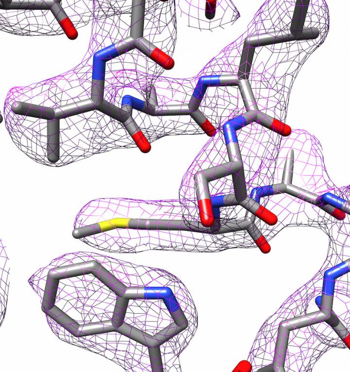
Electron cryo-microscopes are very complex devices. “Even tiny misalignments can render the images useless,” says Kastritis. It is important to programme them correctly and Halle has the technical expertise to do this. But the analysis that is conducted after the data has been collected is just as important. “The microscope produces several thousand images,” explains Kastritis. Image processing programmes are used to create a 3D structure of the molecule. In cooperation with Professor Milton T. Stubbs from the Institute of Biochemistry and Biotechnology at MLU, the researchers have developed a new method to create a high-resolution model of a protein. Stubbs’ research group uses X-ray crystallography, another technique for determining the structure of proteins, which requires the proteins to be crystallised. They were able to combine a modified form of an image analysis technique with the images taken with the electron cryo-microscope. This made charge states and individual water molecules visible.
“It’s an attractive method,” says Kastritis. Instead of needing very expensive microscopes, a lot of computing capacity is required, which MLU has. Now, in addition to using X-ray crystallography, electron cryo-microscopy can be used to produce images of proteins – especially those that are difficult to crystallise. This enables collaboration, both inside and outside the university, on the structural analysis of samples with medical and biotechnological potential.
###
Media Contact
Ronja Münch
[email protected]
Original Source
https:/
Related Journal Article
http://dx.
