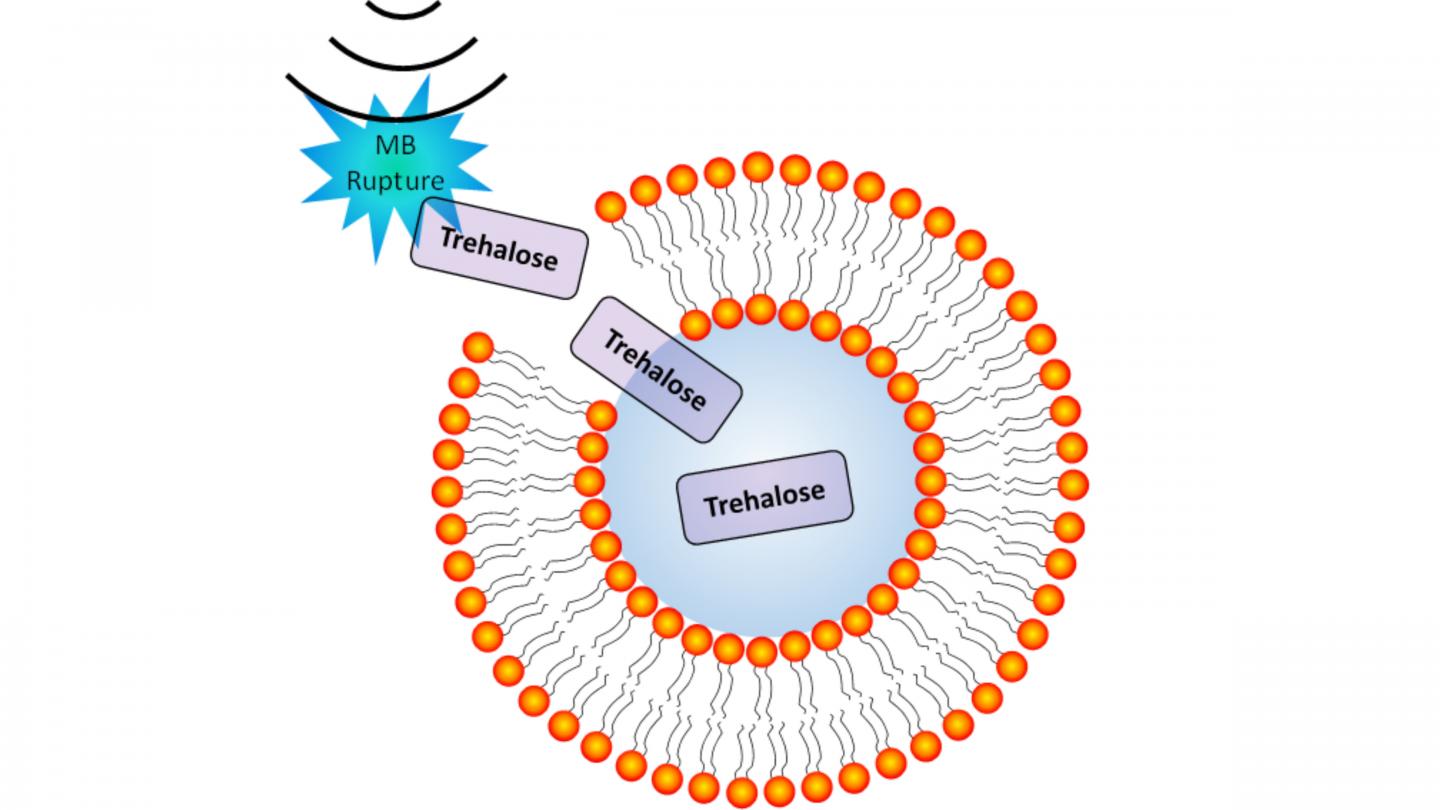
Fine-tuning microbubbles creates pores in blood cells and allows the insertion of molecules, including a special sugar used by some of the hardiest microorganisms to weather extreme environments

Credit: Jonathan A Kopechek
WASHINGTON, April 21, 2020 — Ensuring adequate preservation of the millions of units of blood that are donated every year presents a challenge for blood banks, as blood can typically be stored for only six weeks after donation. A potential solution to the problem attempts to dry blood by using a sugar-based preservative that organisms living in some of Earth’s most extreme environments produce to weather long periods of dryness. New work in ultrasound technology looks to provide a path to inserting these sugars into human red blood cells, in an effort to help them last for years.
Researchers at the University of Louisville have demonstrated a new way to use ultrasound to create pores in blood cells, which allows the molecule trehalose to enter the cells and prevent their degradation when dried for preservation. Oscillating microscopic bubbles of inert gases with ultrasound provides the microfluidic system with the ability to increase the number of viable cells that can be rehydrated. The researchers discuss their work in this week’s Biomicrofluidics, from AIP Publishing.
The approach could lead to ways of increasing the shelf life of blood donations from weeks to the order of years. Such advances would provide a boon to those needing blood in areas where access to donations is difficult, like on the battlefield or in space.
“What’s unique about this is there are not many other studies that look at using acoustofluidics to place a molecule like this inside red blood cells,” said author Jonathan Kopechek. “It’s also interesting, because it allows us to store blood without keeping it cool.”
When it’s not helping these extremophiles survive, trehalose is a relatively cheap sugar that is so safe that it is used as a preservative for food items, like donut glaze.
The group constructed a spiral-shaped channel that exposed blood cells to trehalose while surrounded by microbubbles. They tuned ultrasonic vibrations using several parameters until the bubbles shook nanosized holes in the membranes of the blood cells, just large enough for trehalose or a closely related fluorescent molecule called fluorescein to enter and just briefly enough to maintain the integrity of the blood.
After confirming that fluorescein could enter cells on test samples, they added trehalose to a new batch of samples, dried the blood, rehydrated it, and performed tests to count how many of the blood cells were still viable after the process.
The ultrasound technique was able to preserve a significantly higher portion of cells by adding trehalose versus leaving the trehalose out.
The group looks to improve on the yield for their technique with the hopes of verifying the effectiveness of dry-preserved blood in patients.
###
The article, “Ultrasound-induced molecular delivery to erythrocytes using a microfluidic system,” is authored by Connor Sterling Centner, Emily M. Murphy, Mariah C. Priddy, John T. Moore, Brett R. Janis, Michael A. Menze, Andrew P. DeFilippis and Jonathan A. Kopechek. The article will appear in Biomicrofluidics on April 21, 2020 (DOI: 10.1063/1.5144617). After that date, it can be accessed at https:/
ABOUT THE JOURNAL
Biomicrofluidics rapidly disseminates research in fundamental physicochemical mechanisms associated with microfluidic and nanofluidic phenomena. The journal also publishes research in unique microfluidic and nanofluidic techniques for diagnostic, medical, biological, pharmaceutical, environmental, and chemical applications. See https:/
Media Contact
Larry Frum
[email protected]
Related Journal Article
http://dx.


