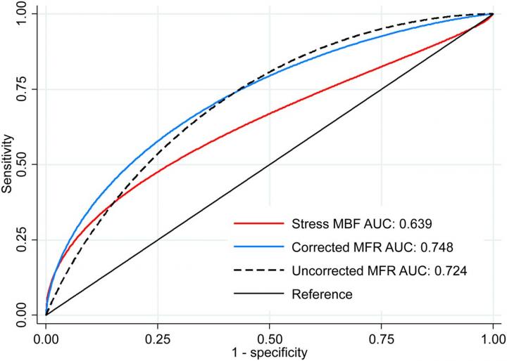
Credit: RJH Miller and O Manabe et al. Departments of Imaging and Medicine, Cedars-Sinai Medical Center, Los Angeles, CA, USA.
Myocardial blood flow (MBF) and myocardial flow reserve (MFR) have been identified as accurate indicators for graft failure after cardiac transplantation, according to a new study published in The Journal of Nuclear Medicine. Utilizing positron emission tomography (PET) myocardial perfusion imaging to quantify MBF and MRF, researchers were able to successfully detect patients with cardiac allograft vasculopathy (CAV), the most serious condition facing transplant patients late after their surgery. In addition, researchers found that MFR had a significantly higher accuracy when predicting the overall prognosis for cardiac transplant patients.
Heart transplantation is a definitive therapy for patients with end-stage heart failure and has a median post-transplant survival of more than 13 years. As long-term survival has increased, the prevalence of cardiac diseases, such as CAV, has also grown. CAV accounts for over one-third of deaths in patients who survive at least five years after their heart transplant. It is also the most common indication for re-transplantation in patients who survive one year.
“MBF and MFR have been shown to be useful for diagnosis and prognosis of CAV in a few single-center studies, however, there is no consensus on which marker–stress MBF or MFR–should be applied for these purposes,” said Robert J.H. Miller, MD, FRCPC, clinical assistant professor at the University of Calgary in Canada. “In this study, we compared the utility of MBF and MFR, using previously derived thresholds, to provide the external validation required to guide broader clinical implementation.”
Ninety-nine cardiac transplant patients who underwent 82Rb PET myocardial perfusion imaging over a five-year period in a single center were included in the study. Quantitative and semi-quantitative analyses were performed and imaging parameters were compared among study participants who died (26 patients) and those who survived (73 patients). Researchers then examined the diagnostic and prognostic accuracy for MBR and MFR in detecting significant CAV.
Results from the study demonstrated that stress MBF, uncorrected MFR and corrected MFR were equivalent in identifying patients with significant CAV. In terms of prognosis, researchers found that uncorrected MFR offered superior discrimination for mortality of all causes compared to stress MBF. Further, the study found that preserved MFR (defined as greater than or equal to 2.0) identified low-risk patients, while the presence of multiple abnormal parameters identified patients at the highest risk.
“PET with routine measures of MBF and MFR has a clear role in patients following cardiac transplantation. This study provides practical information for centers implementing PET for CAV surveillance and will help guide them in implementing these important measurements” noted Miller.
###
This study was made available online in August 2019 ahead of final publication in print in February 2020.
The authors of “Comparative Prognostic and Diagnostic Value of Myocardial Blood Flow and Myocardial Flow Reserve After Cardiac Transplantation” include Robert J.H. Miller, Balaji Tamarappoo, Sean Hayes, John D. Friedman, Piotr J. Slomka and Daniel S. Berman, Department of Imaging and Medicine, Cedars-Sinai Medical Center, Los Angeles, California; Osamu Manabe, Department of Imaging and Medicine, Cedars-Sinai Medical Center, Los Angeles, California, and Department of Nuclear Medicine, Hokkaido University Graduate School of Medicine, Sapporo, Japan; Jignesh Patel and Jon A. Kobashigawa, Smidt Heart Institute, Cedars-Sinai Heart Institute, Cedars-Sinai Medical Center, Los Angeles, California.
Please visit the SNMMI Media Center for more information about molecular imaging and precision imaging. To schedule an interview with the researchers, please contact Rebecca Maxey at (703) 652-6772 or [email protected]. Current and past issues of The Journal of Nuclear Medicine can be found online at http://jnm.
About the Society of Nuclear Medicine and Molecular Imaging
The Journal of Nuclear Medicine (JNM) is the world’s leading nuclear medicine, molecular imaging and theranostics journal, accessed close to 10 million times each year by practitioners around the globe, providing them with the information they need to advance this rapidly expanding field.
JNM is published by the Society of Nuclear Medicine and Molecular Imaging (SNMMI), an international scientific and medical organization dedicated to advancing nuclear medicine and molecular imaging–precision medicine that allows diagnosis and treatment to be tailored to individual patients in order to achieve the best possible outcomes. For more information, visit http://www.
This work was supported in part by the Dr. Miriam and Sheldon Adelson Medical Research Foundation. Robert Miller receives funding support from the Arthur J.E. Child Fellowship grant. Daniel Berman and Piotr Slomka participate in software royalties for QPS software at Cedars-Sinai Medical Center.
Media Contact
Rebecca Maxey
[email protected]
703-652-6772
Original Source
https:/
Related Journal Article
http://dx.


