Credit: Arvind Pathak
Johns Hopkins Medicine researchers have created a computer program for scientists at no charge that lets users readily quantify the structural and functional changes in the blood flow networks feeding tumors.
The researchers published a link to download the new program, called HemoSYS, and an accompanying manual with instructions on how to use it, on Feb. 11 in Scientific Reports.
“Compared to blood flow in healthy tissues, tumor blood flow is abnormal, and these abnormalities can be captured with new imaging methods. Therefore, we created a freely available toolkit called HemoSYS that enables scientists to quantify these abnormalities from imaging data acquired from tumors in live animals”, says Arvind Pathak, Ph.D., associate professor of radiology and biomedical engineering at the Johns Hopkins University School of Medicine and a member of the Johns Hopkins Sidney Kimmel Comprehensive Cancer Center.
Studying the architecture of blood vessels and their flow dynamics in tumors could provide insights into cancer progression and metastasis, says Janaka Senarathna, Ph.D., a research fellow in Pathak’s lab and lead author of the paper. This approach could accelerate development of new therapies that target a tumor’s blood vessels in order to limit its supply of nutrients and oxygen. HemoSYS could also lead to more effective delivery of already available drugs by mapping blood flow fluctuations in the vessels feeding the tumor.
A tumor’s blood vessels are its lifeline for survival and growth, providing it with nutrients as well as an avenue for tumor cells to spread to other parts of the body. However, these vessels often grow irregularly and create abnormal blood flow patterns, making them a huge hurdle to effective delivery of therapeutics.
“The abnormal blood flow makes it difficult to predict how effective therapies will be, and if insufficient drug is delivered to the tumor, cancers may recur or develop resistance to treatment or advance harmful side effects,” says Pathak.
The Johns Hopkins investigators caution that the research tool is not directly applicable to human tumors yet. But, says Pathak, “As our ability to obtain high-resolution images in the clinic improves, we hope that this tool can be adapted to provide a noninvasive way to analyze the blood flow fluctuations in an individual patient’s cancer and help to customize their therapy.”
To develop HemoSYS, the Pathak Lab recruited biomedical engineers and biophysicists to develop accurate, efficient ways to quantify “multivariable” data comprised of tumor blood flow, blood volume and oxygenation images. A different type of light source was used to collect each of these data variables from tumors implanted in animals.
“Typically, a research lab studying these blood vessel systems would need to have extensive expertise in image processing to quantify the relationships between these measurements,” says Pathak. “HemoSYS allows researchers without any programming expertise to conduct their analyses on these multivariable imaging data.”
The HemoSYS program employs fundamental engineering principles to analyze and integrate different kinds of data collected from various imaging methods.
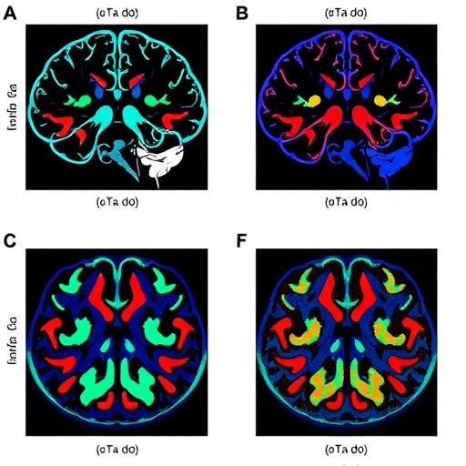
This data enables scientists to rigorously map the entire “hemodynamic landscape” of the tumor being studied, says Pathak. The result is a colorful yet informative visualization that shows the relationships among blood flow, oxygenation and tumor cells in vivid reds, blues and greens.
“The system lets researchers see that there may be an area in the tumor of low oxygen supply caused by poor blood flow, or see that an area is low on oxygen even though it’s exhibiting elevated blood volume,” says Pathak.
Scientists and clinicians can download the toolkit at pathaklab.org/Hemosys. Pathak and his team have also made a customizable version for those who want to adapt HemoSYS for their own experiments or for data acquired with other imaging methods such as MRI, ultrasound and dynamic CT.
###
Other researchers involved in this study include Ayush Prasad, Akanksha Bhargava, Stacy Gil and Nitish Thakor of the Johns Hopkins University School of Medicine.
This work was supported by the National Cancer Institute (1R01CA196701, 5R01CA138264) and a Kavli Neuroscience Distinguished Fellowship.
The authors declare no competing interests.
Media Contact
Rachel Butch
[email protected]
410-955-8665
Original Source
https:/

