• HOME
  • NEWS
  • EXPLORE
    • CAREER
      • Companies
      • Jobs
    • EVENTS
    • iGEM
      • News
      • Team
    • PHOTOS
    • VIDEO
    • WIKI
  • BLOG
  • COMMUNITY
    • FACEBOOK
    • INSTAGRAM
    • TWITTER
Sunday, August 16, 2026
BIOENGINEER.ORG
No Result
View All Result
  • Login
  • HOME
  • NEWS
  • EXPLORE
    • CAREER
      • Companies
      • Jobs
        • Lecturer
        • PhD Studentship
        • Postdoc
        • Research Assistant
    • EVENTS
    • iGEM
      • News
      • Team
    • PHOTOS
    • VIDEO
    • WIKI
  • BLOG
  • COMMUNITY
    • FACEBOOK
    • INSTAGRAM
    • TWITTER
  • HOME
  • NEWS
  • EXPLORE
    • CAREER
      • Companies
      • Jobs
        • Lecturer
        • PhD Studentship
        • Postdoc
        • Research Assistant
    • EVENTS
    • iGEM
      • News
      • Team
    • PHOTOS
    • VIDEO
    • WIKI
  • BLOG
  • COMMUNITY
    • FACEBOOK
    • INSTAGRAM
    • TWITTER
No Result
View All Result
Bioengineer.org
No Result
View All Result
Home NEWS Science News Health

New approach uses light to stabilize proteins for study

Bioengineer by Bioengineer
November 4, 2019
in Health
Reading Time: 3 mins read
0
IMAGE
Share on FacebookShare on TwitterShare on LinkedinShare on RedditShare on Telegram

IMAGE

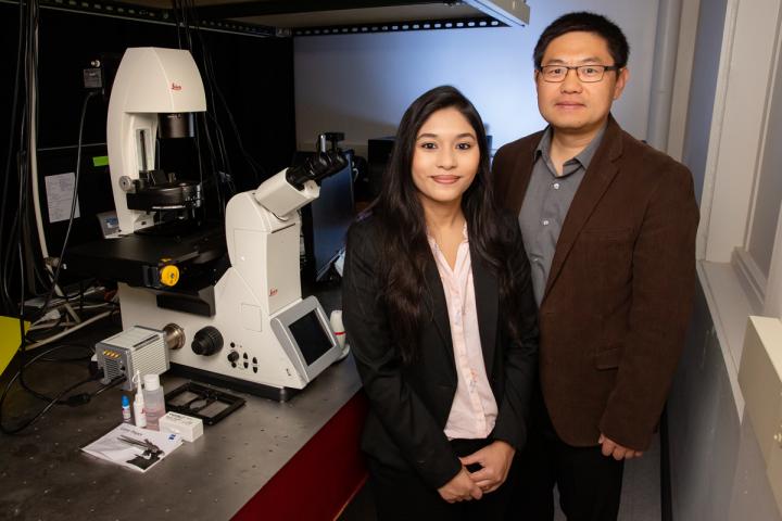
Credit: Photo by L. Brian Stauffer


CHAMPAIGN, Ill. – Researchers report they have developed a new technique that uses light to control the lifetime of a protein inside the cell. This method will allow scientists to better observe how specific proteins contribute to health, development and disease.

Previous techniques for controlling protein levels involved adding chemicals that degrade specific proteins, said University of Illinois biochemistry professor Kai Zhang, who led the new research. Using light, a method called optogenetics, is a more efficient, nontoxic way to control protein levels, Zhang said.

In earlier optogenetics approaches, scientists modified specific proteins so that shining a light on the cell caused the proteins to break down, Zhang said.

“We wanted to develop a system where you can stabilize – rather than degrade – a target protein using light,” said Payel Mondal, a graduate student in the Zhang lab. “This can have applications in cancer where you want to stabilize a cancer inhibitor.”

Proteins are activated in cells in different ways. The researchers wanted to ensure that their method would work on any protein of interest.

The team’s new technique, called GLIMPSe, involves attaching a short peptide sequence, called a degron, to the target protein that signals the cell to degrade it. Light triggers the cell to remove the degron, thus rescuing the protein from degradation. This technique allows scientists to study what happens when a protein is present or absent in the cell, or when it is present at lower and higher levels.

The researchers modified two kinds of proteins: a kinase and a phosphatase.

“If you activate the kinase, it will direct the cell to differentiate into a neuronal cell line,” Zhang said. “If you activate the phosphatase, it will block that differentiation.”

“We demonstrated that the stability of two different classes of proteins can be controlled using light,” Mondal said. “We saw protein stabilization within 30 minutes of using the light.”

“One of the limitations of this system is that once the protein is rescued, we have no further ability to control its levels,” Zhang said. “Eventually, the cell will degrade the protein.”

The researchers are working to develop new techniques to further extend their control.

The findings are the result of a long-term collaboration with Jing Yang, a University of Illinois comparative biosciences professor and a co-author of the paper.

“We started looking at embryonic development and Professor Yang had a very detailed study of how the degron worked,” Zhang said. “By bridging embryonic development with synthetic biology, we created new tools.”

Zhang’s team is studying how to control protein stability after its optogenetic rescue and how to use this technique to study embryonic development.

The new findings are reported ACS Synthetic Biology.

###

The National Institutes of Health supported this work.

Editor’s notes:

To reach Kai Zhang, call 217-300-0582; email [email protected].

The paper “Repurposing protein degradation for optogenetic modulation of protein activities” is available online and from the U. of I. News Bureau.

Media Contact
Ananya Sen
[email protected]
217-333-5802

Original Source

https://news.illinois.edu/view/6367/804107

Related Journal Article

http://dx.doi.org/10.1021/acssynbio.9b00285

Tags: BiochemistryCell BiologyMedicine/HealthneurobiologyPharmaceutical SciencePharmaceutical Sciences
Share12Tweet8Share2ShareShareShare2

Related Posts

PARP1 Drives Neuropathic Pain Through GPX4-Dependent Ferroptosis in Injured Mice’s Sensory Neurons

August 15, 2026

Genome-wide skin pQTL map reveals genetic regulators and mechanisms underlying skin disorders

August 15, 2026

Levistilide A Drives Ferroptosis via RNF40-HSP90α Axis, Suppressing Colorectal Cancer Lung Metastasis

August 15, 2026

Sintilimab, IBI305, and Chemotherapy Show Promise in First-Line Advanced Gastric and GEJ Adenocarcinoma

August 15, 2026
Please login to join discussion

POPULAR NEWS

  • KAIST develops semiconductor neuron that harnesses noise to selectively process signals

    29 shares
    Share 12 Tweet 7
  • Imagining natural and extra robotic thumbs together strengthens kinesthetic sensorimotor networks

    29 shares
    Share 12 Tweet 7
  • PARP1 Drives Neuropathic Pain Through GPX4-Dependent Ferroptosis in Injured Mice’s Sensory Neurons

    29 shares
    Share 12 Tweet 7
  • Strubbelig–NHL3 Receptor Complex Helps Arabidopsis Respond to Cellulose Deficiency

    29 shares
    Share 12 Tweet 7

About

We bring you the latest biotechnology news from best research centers and universities around the world. Check our website.

Follow us

Recent News

KAIST develops semiconductor neuron that harnesses noise to selectively process signals

Imagining natural and extra robotic thumbs together strengthens kinesthetic sensorimotor networks

PARP1 Drives Neuropathic Pain Through GPX4-Dependent Ferroptosis in Injured Mice’s Sensory Neurons

Subscribe to Blog via Email

Success! An email was just sent to confirm your subscription. Please find the email now and click 'Confirm' to start subscribing.

Join 85 other subscribers
  • Contact Us

Bioengineer.org © Copyright 2023 All Rights Reserved.

Welcome Back!

Login to your account below

Forgotten Password?

Retrieve your password

Please enter your username or email address to reset your password.

Log In
No Result
View All Result
  • Homepages
    • Home Page 1
    • Home Page 2
  • News
  • National
  • Business
  • Health
  • Lifestyle
  • Science

Bioengineer.org © Copyright 2023 All Rights Reserved.