Researchers try a trick for complete 3D analysis of submicron crystals
Credit: © Tim Grüne
The 3D analysis of crystal structures requires a full 3D view of the crystals. Crystals as small as powder, with edges less than one micrometer, can only be analysed with electron radiation. With electron crystallography, a full 360-degree view of a single crystal is technically impossible. A team of researchers led by Tim Gruene from the Faculty of Chemistry at the University of Vienna modified the holder of the tiny crystals so that a full view becomes possible. Now they presented their solutions in the journal „Nature Communications”.
Typically, crystallographers use X-rays to examine their samples. Size, however, matters greatly for X-ray structure analysis: Crystals with edges less than 50 to 100 micrometres are too small to produce a measurable signal. “Electron crystallography is a quite recent development. We demonstrated to our chemist colleagues that we can analyse crystals with edges less than 1 micrometre – this includes many crystals which escape 3D structure determination so far”, Tim Grüne says, who is member of the Department of Inorganic Chemistry and head of the Centre for X-ray Structure Analysis.
Limited View
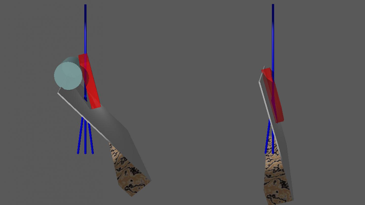
Electrons interact with matter much stronger than X-rays. Submicrometre sized crystals produce characteristic diffraction images when they are irradiated with electrons. These provide the data for structure analysis. However, the sample holder prevents a full 360 degree rotation: Currently only one rotation axis is available, and the metal bars necessary to stabilise the delicate cannot be penetrated by the electrons. Only a rotation of about 75 degrees is possible in either direction. „This gives us a maximum of 300 degree valuable data, which leads to an erroneous structural analysis”, says Gruene. He and his colleagues from ETH Zurich and from PSI came up with a neat trick to solve the problem.
Their study presents two solutions to circumvent the problem: They prepared the sample holder so that crystals can be viewed from all sides. One sample holder contains dozens of crystals, more than enough to complete the data and provide an undistorted 3D view.
Tricking the Carrier
A simple, readily available means disturbs the carrier material, an ultrathin carbon layer, with a fine brush. According to Gruene “as a consequence, individual segments of the carbon layer curl up – like when you touch the fruit of touch-me-not. The crystals stick to the curls and achieve a random orientation. One can comfortably select several individual crystals from very different views”.
The second solution covers the carbon carrier with nylon fibres. „The surfaces resembles a forest covered chaotically with tree logs”, Tim Grüne says. This again leads to many random orientations of the crystals when they are deposited on the sample holder. However, the nylon fibres are deposited with electrospinning, which requires an additional apparatus and is a bit more complex than stroking it with a brush.
“Neat and simple”
Both measures provide data sets from the crystals with a complete 3D structural analysis. This type of combining data sets is common practice in protein crystallography, but much less common in chemical crystallography. Tim Grüne explains, “Our work exploited the fact that data merging works likewise for chemical compounds as it does for proteins. We only needed 5 crystals in both cases to complete the data”.
„We did not avoid the problem, but demonstrated how to reveal the hidden faces of the crystals to the electron beam. Both solutions are surprisingly simple and can be realised without much effort”, says Tim Grüne.
###
Publication in Nature Communications:
“3D-structured supports create complete data sets for electron crystallography” J. T. C. Wennmacher, C. Zaubitzer, T. Li, Y. K. Bahk, J. Wang, J. A. van Bokhoven, T. Gruene, Nature Communications, 25 July 2019.
DOI: 10.1038/s41467-019-11326-2
Media Contact
Tim Grüne
[email protected]
Original Source
https:/
Related Journal Article
http://dx.


