Credit: Carnegie Mellon University College of Engineering
PITTSBURGH–Carnegie Mellon University’s Assistant Professor of Electrical and Computer Engineering (ECE) Maysam Chamanzar and ECE Ph.D. student Matteo Giuseppe Scopelliti today published research that introduces a novel technique which uses ultrasound to noninvasively take optical images through a turbid medium such as biological tissue to image body’s organs. This new method has the potential to eliminate the need for invasive visual exams using endoscopic cameras.
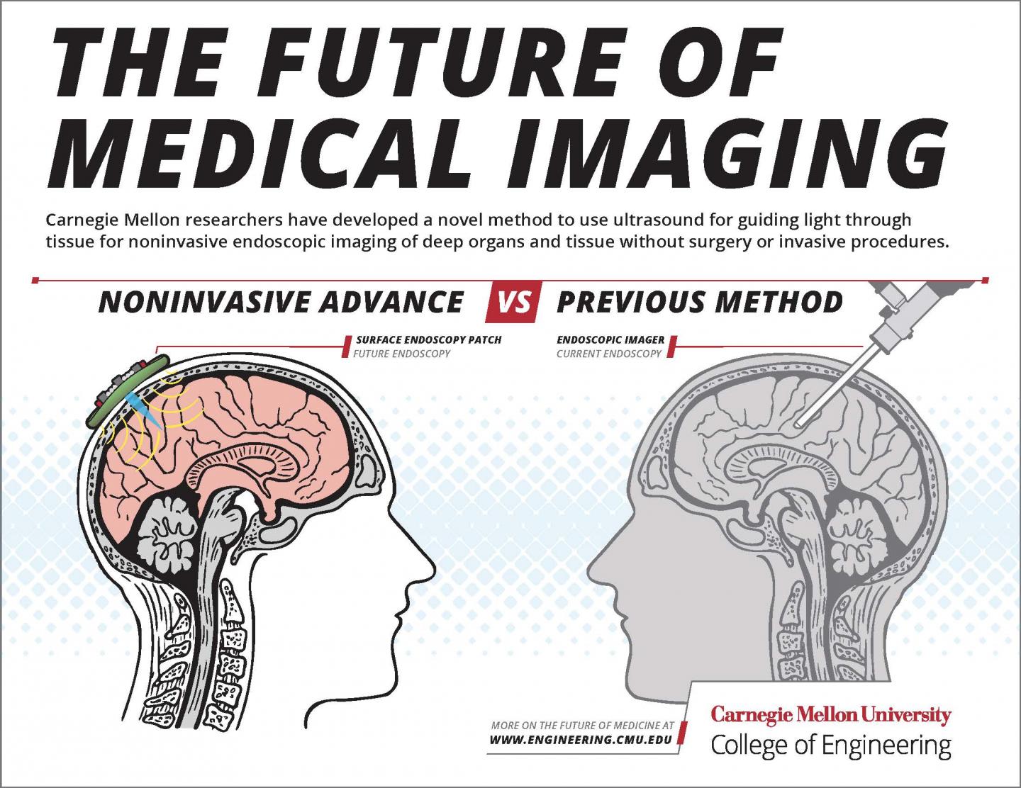
In other words: one day, scopes may no longer need to be inserted into the body, such as down the throat or under the skin, to reach the stomach, brain, or any other organs for examination.
Endoscopic imaging, or using cameras inserted directly inside the body’s organs to investigate symptoms, is an invasive procedure used to examine and diagnose symptoms of deep tissue disease. Endoscopic imagers, or cameras on the end of catheter tubes or wires, are usually implanted through a medical procedure or surgery in order to reach the body’s deep tissues, but Chamanzar’s new technique provides a completely non-surgical and noninvasive alternative.
The lab’s paper published in Light: Science and Applications, a journal published by Springer Nature, shows that they can use ultrasound to create a virtual “lens” within the body, rather than implanting a physical lens. By using ultrasonic wave patterns, the researchers can effectively “focus” light within the tissue, which allows them to take images never before accessible through noninvasive means.
Biological tissue is able to block most light, especially light in the visible range of the optical spectrum. Therefore, current optical imaging methods cannot use light to access deep tissue from the surface. Chamanzar’s lab, however, has used noninvasive ultrasound to induce more transparency to enable more penetration of light through turbid media, such as biological tissue.
“Being able to relay images from organs such as the brain without the need to insert physical optical components will provide an important alternative to implanting invasive endoscopes in the body,” says Chamanzar. “We used ultrasound waves to sculpt a virtual optical relay lens within a given target medium, which for example, can be biological tissue. Therefore, the tissue is turned into a lens that helps us capture and relay the images of deeper structures. This method can revolutionize the field of biomedical imaging.”
Ultrasound waves are able to compress and rarefy, or thin, whatever medium they are flowing through. In compressed regions, light travels more slowly compared to rarefied regions. In this paper, the team shows that this compression and rarefication effect can be used to sculpt a virtual lens in the target medium for optical imaging. This virtual lens can be moved around without disturbing the medium simply by reconfiguring the ultrasound waves from outside. This enables imaging different target regions, all noninvasively.
The published method is a platform technology that can be applied in many different applications. In future, it can be implemented in the form of a handheld device or wearable surface patch, depending on the organ being imaged. By placing the device or patch on the skin, the clinician would be able to easily receive optical information from within the tissue to create images of what’s inside without endoscopy’s many discomforts and side effects.
The closest current applications for this technology would be endoscopic imaging of brain tissue or imaging under the skin, but this technique can also be used in other parts of the body for imaging. Beyond biomedical applications, this technique can be used for optical imaging in machine vision, metrology, and other industrial applications to enable non-destructive and steerable imaging of objects and structures at the micron scale.
The researchers showed that the properties of the virtual “lens” can be tuned by changing the parameters of the ultrasonic waves, allowing users to “focus” images taken using the method at different depths through the medium. While the LSA paper is focused on the method’s efficacy for closer-to-the-surface applications, the team has yet to find the limit to how deep within the body’s tissue this ultrasonically-assisted optical imaging method can reach.
“What distinguishes our work from conventional acousto-optic methods is that we are using the target medium itself, which can be biological tissue, to affect light as it propagates through the medium,” explains Chamanzar. “This in situ interaction provides opportunities to counterbalance the non-idealities that disturb the trajectory of light.”
This technique has many potential clinical applications, such as diagnosing skin disease, monitoring brain activity, and diagnosis and photodynamic therapy for identifying and targeting malignant tumors.
In addition to the direct implications this research has on clinical medicine, it will also have indirect clinical applications. By using this acousto-optic technology to view mouse models of brain disorders in action and selectively stimulate different neural pathways, researchers would be able to study the mechanisms involved in disease conditions such as Parkinson’s, informing the design of next generation clinical therapeutic interventions to treat these diseases in humans.
“Turbid media have always been considered obstacles for optical imaging,” says Scopelliti. “But we have shown that such media can be converted to allies to help light reach the desired target. When we activate ultrasound with the proper pattern, the turbid medium becomes immediately transparent. It is exciting to think about the potential impact of this method on a wide range of fields, from biomedical applications to computer vision.”
###
The researchers project that this new imaging technology could be applied in biomedical and clinical contexts within the next five years.
About the College of Engineering: The College of Engineering at Carnegie Mellon University is a top-ranked engineering college that is known for our intentional focus on cross-disciplinary collaboration in research. The College is well-known for working on problems of both scientific and practical importance. Our “maker” culture is ingrained in all that we do, leading to novel approaches and transformative results. Our acclaimed faculty have a focus on innovation management and engineering to yield transformative results that will drive the intellectual and economic vitality of our community, nation and world.
About Carnegie Mellon University: Carnegie Mellon is a private, internationally ranked university with programs in areas ranging from science, technology and business to public policy, the humanities and the arts. More than 13,000 students in the university’s seven schools and colleges benefit from a small faculty-to-student ratio and an education characterized by its focus on creating and implementing solutions for real world problems, interdisciplinary collaboration and innovation.
Media Contact
Emily Durham
[email protected]
Original Source
https:/

