• HOME
  • NEWS
  • EXPLORE
    • CAREER
      • Companies
      • Jobs
    • EVENTS
    • iGEM
      • News
      • Team
    • PHOTOS
    • VIDEO
    • WIKI
  • BLOG
  • COMMUNITY
    • FACEBOOK
    • INSTAGRAM
    • TWITTER
Monday, August 25, 2025
BIOENGINEER.ORG
No Result
View All Result
  • Login
  • HOME
  • NEWS
  • EXPLORE
    • CAREER
      • Companies
      • Jobs
        • Lecturer
        • PhD Studentship
        • Postdoc
        • Research Assistant
    • EVENTS
    • iGEM
      • News
      • Team
    • PHOTOS
    • VIDEO
    • WIKI
  • BLOG
  • COMMUNITY
    • FACEBOOK
    • INSTAGRAM
    • TWITTER
No Result
View All Result
Bioengineer.org
No Result
View All Result
Home NEWS Science News Science

Speedy ‘slingshot’ cell movement observed for the first time

Bioengineer by Bioengineer
March 12, 2019
in Science
Reading Time: 2 mins read
0
Share on FacebookShare on TwitterShare on LinkedinShare on RedditShare on Telegram

ANN ARBOR–By slingshotting themselves forward, human cells can travel more than five times faster than previously documented.

University of Michigan researchers observed the movement in bioengineered 3D scaffolds that model stromal tissue–the connective tissue that surrounds organs.

The researchers say this method of cell movement, observed for the first time, could be involved in the spread of cancer. And in the future it could potentially be harnessed to direct the movement of healthy cells for tissue repair therapies.

The cells pull on and stretch the surrounding fibrous tissue, eventually using that tension to launch themselves forward. The motion looked familiar enough that researchers dubbed it “slingshot migration.”

“We found that cells can move in this very distinct way that results in effective migration far faster than anything previously reported,” said Brendon Baker, U-M assistant professor of biomedical engineering.

William Wang, a doctoral student in biomedical engineering, was the first to witness it. He was studying the properties of stromal tissue that either hinder or promote the spread of cells in diseases such as cancer.

“I was definitely shocked seeing a cell move so fast,” Wang said. “What was even more surprising was then capturing this migration mode in multiple cell types and finding that their speed was so much faster than traditional modes of cell migration.”

Research in this area typically involves observing cells under the microscope on a flat petri dish. But that doesn’t provide a complete picture.

“These flat, two-dimensional surfaces really don’t resemble the structure and mechanical behavior of real tissues, especially the ones where so much of the migration in our body is occurring,” Wang said.

The researchers study cells in a 3D fibrous environment, which is closer to the real thing.

Most organs can be broken down into two parts: functional elements such as sacs, ducts or glands called the parenchyma, and the surrounding collagenous tissue that supports the blood vessel network, called the stroma or interstitia.

Baker’s lab builds stromal tissue for two applications: to study how cells behave during disease progression, and to advance organ replacement therapies.

Cancer cells that remain within the parenchyma, where most originate, can typically be dealt with effectively by surgical removal. When they migrate into the stromal tissue, they pose a much greater threat.

“It’s metastasis, or the spread of the cancer, that kills patients,” Baker said. “For that to happen, the cells have to break out of the parenchyma and cross through the fibrous stroma to reach blood or lymphatic vessels or the lymphatic system.”

###

The research, published in Nature Communications, is funded by the National Institutes of Health.

Study: Actomyosin contractility-dependent matrix stretch and recoil induces rapid cell migration

Brendon Baker

Media Contact
Nicole Moore

[email protected]

Tags: BiologyMedicine/HealthTechnology/Engineering/Computer Science
Share12Tweet7Share2ShareShareShare1

Related Posts

Five or more hours of smartphone usage per day may increase obesity

July 25, 2019
IMAGE

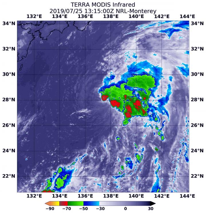
NASA’s terra satellite finds tropical storm 07W’s strength on the side

July 25, 2019

NASA finds one burst of energy in weakening Depression Dalila

July 25, 2019

Researcher’s innovative flood mapping helps water and emergency management officials

July 25, 2019
Please login to join discussion

POPULAR NEWS

  • blank

    Molecules in Focus: Capturing the Timeless Dance of Particles

    142 shares
    Share 57 Tweet 36
  • Breakthrough in Computer Hardware Advances Solves Complex Optimization Challenges

    141 shares
    Share 56 Tweet 35
  • New Drug Formulation Transforms Intravenous Treatments into Rapid Injections

    115 shares
    Share 46 Tweet 29
  • Neuropsychiatric Risks Linked to COVID-19 Revealed

    81 shares
    Share 32 Tweet 20

About

We bring you the latest biotechnology news from best research centers and universities around the world. Check our website.

Follow us

Recent News

Linking Surrogate Endpoints to Outcomes in IgA Nephropathy

In Vivo Insights into Aggregation-Induced Emission

Innovative 3D-Printed Scaffolds Pave the Way for Spinal Cord Injury Recovery

  • Contact Us

Bioengineer.org © Copyright 2023 All Rights Reserved.

Welcome Back!

Login to your account below

Forgotten Password?

Retrieve your password

Please enter your username or email address to reset your password.

Log In
No Result
View All Result
  • Homepages
    • Home Page 1
    • Home Page 2
  • News
  • National
  • Business
  • Health
  • Lifestyle
  • Science

Bioengineer.org © Copyright 2023 All Rights Reserved.