Credit: J.R.Daban
A new study based on electron microscopy techniques at low temperatures demonstrates that, during mitosis, chromosome DNA is packed in stacked layers of chromatin. The research, published in EMBO Journal, confirms a surprising structure proposed by UAB researchers over a decade ago, but criticized due to the limitations of the technique used.
In the cell nuclei the DNA is bound to histone proteins and forms long chains of nucleosomes that are called chromatin fibers. In the Chromatin Laboratory at the Department of Biochemistry and Molecular Biology of the UAB, directed by Professor Joan-Ramon Daban, it was discovered in 2005 that the chromatin of mitotic chromosomes forms multilaminar plates. This was a surprising result, which has been criticized because it was not expected that linear fibers of chromatin could give rise to planar structures, and because it is based on conventional electron microscopy and atomic force microscopy techniques that require adsorbing the sample, respectively, on flat surfaces of carbon and mica. In addition, in the case of electron microscopy, the sample has to be fixed with chemical crosslinkers, treated with contrasting agents, and dehydrated.
A new study based on electron microscopy under cryogenic conditions, and synchrotron X-ray scattering, published in EMBO Journal, has shown that in mitotic chromosomes the DNA is densely packed forming stacked sheets of chromatin, which are stabilized by interactions between nucleosomes.
The advantage of the cryo-electron microscopy techniques, used in this new study, is that the sample (uncrosslinked and untreated with contrasting agents) is suspended in an aqueous solution that is kept frozen at -180 ° C, even during imaging. Since the structures to be studied are large and complex, in this work cryo-electron tomography was used because this technique allows capturing many images with different tilt angles and, in the end, a three-dimensional reconstruction of the analyzed structures is obtained.
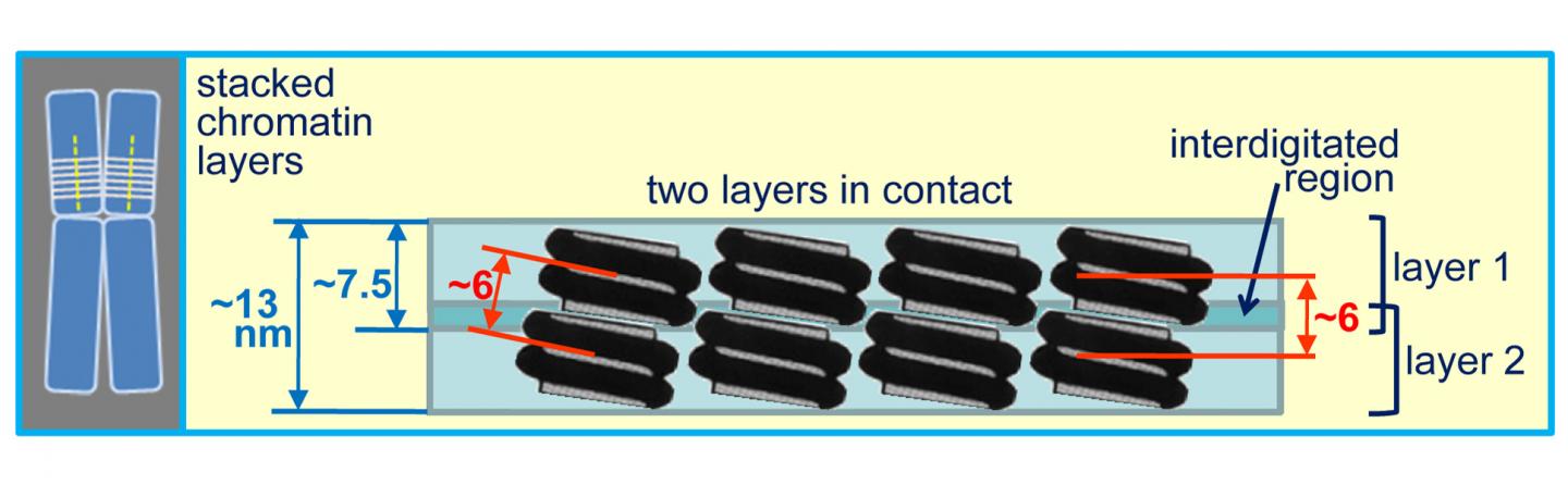
The three-dimensional reconstructions showed that the chromatin emanating from human chromosomes maintained under physiological ionic conditions is planar and forms multilaminar plates. The thickness measurements obtained (single layer 7.5 nm; two layers in contact 13 nm) suggest that the plates are formed by mononucleosome layers, which are interdigitated between them. The complementary X-ray scattering experiments showed a dominant peak at 6 nm, which can be correlated with the distance between layers and between nucleosomes associated through their lateral faces.
There are multilaminar plates that have the dimensions corresponding to the diameter of a human chromosome (600 nm). This suggests that the chromosomes are formed by stacked layers of chromatin that are oriented perpendicular to the axis of the chromosome. This structure is very compact and probably has the function of protecting the integrity of genomic DNA during cell division.
###
The research has been led by the Chromatin Laboratory of the UAB (Andrea Chicano, Eva Crosas, and Joan-Ramon Daban). The small-angle X-ray scattering experiments were performed at the beamline NCD-BL11 of the ALBA synchrotron in Cerdanyola del Vallès. The cryotomograms were obtained with the collaboration of Benjamin Engel in the Instruct (EU) cryo-electron microscopy platform at Max-Planck-Institute of Biochemistry in Martinsried (Germany), and the three-dimensional reconstructions were obtained with the collaboration of Joaquín Otón and Roberto Melero at the Instruct image processing platform at the National Center of Biotechnology in Madrid.
Media Contact
Joan-Ramon Daban
[email protected]
Related Journal Article
http://dx.


