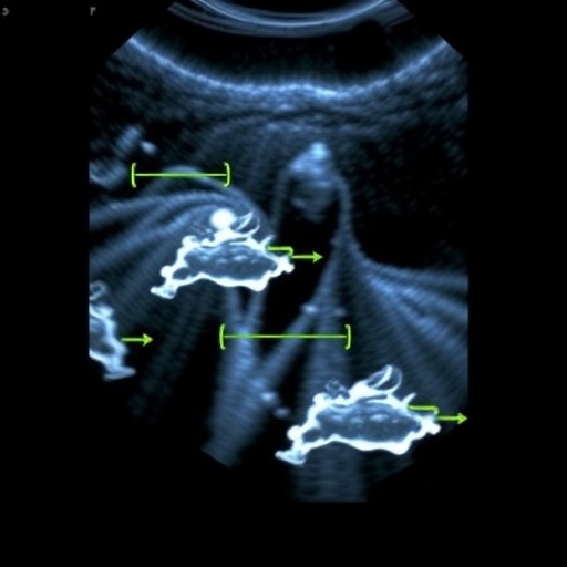

In recent years, the landscape of prenatal diagnosis has undergone a significant transformation, greatly enhancing the ability of healthcare professionals to detect and understand various congenital anomalies before birth. Among the cutting-edge technologies reshaping this field is four-dimensional (4D) direct volume rendering fetal echocardiography, a technique that provides detailed, live images of the fetal heart. Recent studies, including one led by Karmegaraj and Vijayakumar, highlight the potential of this technology in diagnosing conditions such as isolated anomalous course of the left brachiocephalic vein. This research presents a pioneering exploration into the implications of these advanced imaging techniques on pregnancy management and postnatal outcomes.
One of the key advancements in fetal echocardiography is the transition from two-dimensional (2D) static images to a more dynamic 4D rendering approach. The 4D echocardiography allows for real-time visualization, providing an almost tactile sense of the structures within the developing fetal heart. As healthcare providers navigate this tool, they gain insights that were previously obscured, enabling more accurate assessments of congenital anomalies. This is particularly vital since early detection can lead to timely interventions, which are crucial for improving outcomes.
The anomalous course of the left brachiocephalic vein is just one of many cardiac anomalies that can now be identified before birth. This condition, while relatively rare, can lead to significant complications if not recognized and managed appropriately. The innovative strides in echocardiographic technology have enhanced the clarity with which such conditions can be diagnosed. For example, enhanced imaging capabilities allow clinicians to differentiate between normal and anomalous anatomical structures with unprecedented precision, thereby refining the overarching approach to prenatal care.
.adsslot_49Ly2rvcVZ{width:728px !important;height:90px !important;}
@media(max-width:1199px){ .adsslot_49Ly2rvcVZ{width:468px !important;height:60px !important;}
}
@media(max-width:767px){ .adsslot_49Ly2rvcVZ{width:320px !important;height:50px !important;}
}
ADVERTISEMENT
Moreover, the implications of early detection cannot be overstated. Early diagnosis allows families and healthcare teams to prepare for potential challenges in advance. Parents can be adequately informed about the condition, and appropriate, orchestrated care plans can be designed, which may include surgical interventions soon after birth. The psychological and emotional ramifications for families are also significant, as they can feel more empowered when armed with knowledge regarding their baby’s health.
In this rapidly evolving space, the importance of interdisciplinary collaboration cannot be ignored. By pooling expertise from maternal-fetal medicine specialists, pediatric cardiologists, and radiologists, the potential for enhanced outcomes increases exponentially. The melding of various specialties ensures that all angles of patient care are addressed. Such comprehensive approaches can facilitate the successful management of pregnancies complicated by fetal anomalies.
The role of patient education in this context also deserves emphasis. In an era where information is readily available at the click of a button, it’s paramount that expectant parents receive clear, accurate information regarding their unborn child’s health. This necessitates that healthcare providers develop streamlined communication strategies, ensuring that parents understand the implications of various diagnoses. The integration of technology also plays a significant role; providing visual representations of a diagnosis, such as through 4D echocardiography, can enhance understanding and retention.
Beyond immediate prenatal care, the long-term implications of these diagnoses are worth considering. Postnatal outcomes can vary widely based on how well anomalies are addressed before and after birth. Anomalies identified prenatally allow for a multi-faceted approach to postnatal intervention, which might include surgeries, therapies, and other forms of ongoing medical care. Tracking these patients longitudinally can inform future practices and technologies, forming a critical feedback loop in medical research.
The ethical considerations surrounding prenatal diagnosis are also increasingly at the forefront of medical discussions. With the advent of advanced imaging comes the responsibility to ensure that expectant parents are making informed choices about their care. Conversations around quality of life, potential consequences of interventions, and the uncertainties of congenital anomalies must be handled with sensitivity and clarity. As clinicians continue to navigate these discussions, it becomes crucial to balance hope with realistic outcomes.
In conclusion, the convergence of advanced imaging techniques, interdisciplinary collaboration, and a commitment to ethical patient care forms the backbone of modern prenatal diagnostics. As technology continues to evolve, the potential for improved diagnosis and outcomes will only grow, highlighting the importance of ongoing research and development in this vital area of medicine. The ongoing studies shed light on the significance of understanding fetal cardiac conditions, ultimately working towards a brighter future for the next generation.
As prenatal imaging technologies such as echocardiography continue to hit new milestones, researchers like Karmegaraj and Vijayakumar are at the forefront, paving the way for future innovations. The growing body of evidence supporting the efficacy of these tools highlights their role in transforming prenatal care and, ultimately, enhancing the lives of countless children and their families.
Subject of Research: Prenatal diagnosis using four-dimensional fetal echocardiography for isolated anomalous course of left brachiocephalic vein.
Article Title: Prenatal diagnosis conventional/four-dimensional direct volume rendering fetal echocardiography pregnancy and postnatal outcomes of isolated anomalous course of left brachiocephalic vein.
Article References:
Karmegaraj, B., Vijayakumar, S. Prenatal diagnosis conventional/four-dimensional direct volume rendering fetal echocardiography pregnancy and postnatal outcomes of isolated anomalous course of left brachiocephalic vein. Pediatr Radiol (2025). https://doi.org/10.1007/s00247-025-06367-3
Image Credits: AI Generated
DOI: https://doi.org/10.1007/s00247-025-06367-3
Keywords: prenatal diagnosis, fetal echocardiography, four-dimensional imaging, congenital anomalies, brachiocephalic vein, maternal-fetal medicine, patient outcomes, interdisciplinary care, ethical considerations.
Tags: 4D fetal echocardiographyadvanced imaging techniques in obstetricsbrachiocephalic vein anomaliesdynamic imaging in fetal cardiologyearly detection of heart defectsenhancing prenatal care with 4D imagingimplications for pregnancy managementinnovations in fetal echocardiographyisolated anomalous course of left brachiocephalic veinpostnatal outcomes of cardiac anomaliesprenatal diagnosis of congenital anomaliesreal-time fetal heart visualization


