In the realm of pediatric cardiology and congenital heart defect management, the advent of advanced imaging techniques has revolutionized how practitioners visualize and quantify cardiac structures. Among these modalities, cardiac computed tomography (CT) has gained prominence, particularly due to its precision in depicting complex anatomical arrangements inherent in congenital heart disease. The study by Goo and Abdul Latiff delves into a meticulous methodology employed in cardiac CT ventricular volumetry, which offers an essential framework for cardiac evaluation in congenitally afflicted patients.
Cardiac CT volumetry is a sophisticated procedure that utilizes three-dimensional (3D) imaging to assess chamber volumes, thus aiding in the diagnosis and management of pediatric patients with congenital heart anomalies. This imaging technique not only enhances visualization of the heart’s anatomy but also allows for a quantifiable analysis of ventricular volumes that can be critical in therapeutic decision-making. The streamlined approach proposed by the authors serves to demystify the complexities involved in volumetric measurements, equipping clinicians with a valuable tool in their ongoing battle against congenital heart diseases.
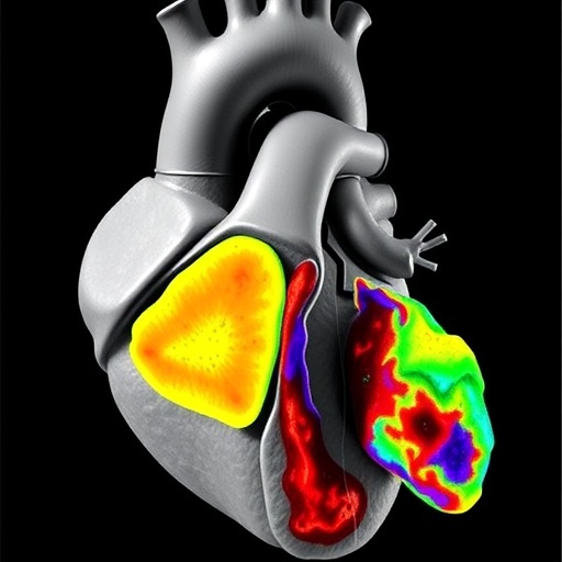
One of the prominent advantages of using cardiac CT is its ability to generate high-resolution images that convey intricate details about the heart’s structure. The methodology outlined by Goo and Abdul Latiff emphasizes a step-by-step guide that incorporates 3D threshold-based segmentation techniques. This technique is particularly significant as it enables an accurate delineation of the heart chambers, ultimately facilitating better assessment of ventricular function and morphology. In pediatric populations where traditional imaging may pose challenges, CT offers an alternative that combines efficiency and remarkable detail.
The procedural steps initiated in this guide underscore the importance of meticulousness in image acquisition and post-processing. To achieve reliable volumetric results, the authors recommend specific preparatory measures, including optimal patient positioning and the careful selection of imaging protocols. These foundational steps are crucial, as they influence the overall quality and diagnostic utility of the images obtained. Emphasizing patient comfort and safety while ensuring adherence to radiation dose optimization principles is paramount in pediatric cardiac imaging.
Segmentation, the heart of the volumetric analysis, is where innovative software truly shines. The authors highlight software tools capable of automating the delineation of cardiac structures, which not only increases reproducibility but also enhances workflow efficiency. As congenital heart disease often necessitates regular monitoring and assessment, the implementation of automated techniques can significantly reduce the time and effort involved in volumetric calculations, ensuring that medical professionals can focus on patient care and interpretation of results.
Moreover, this 3D threshold-based segmentation approach offers invaluable insights beyond pure volumetric data. By allowing for the visualization of dynamic changes in ventricular volume during the cardiac cycle, the methodology enriches our understanding of ventricular function and can gauge the effectiveness of therapeutic interventions. Recognizing the intricate interplay between anatomy and function is vital for the management of patients with congenital heart defects, and enhanced imaging techniques can provide the requisite data for informed clinical decisions.
As technology continues to advance, the future of cardiac imaging suggests even more improvements in the precision of volumetric assessments. The integration of artificial intelligence (AI) and machine learning into cardiac CT volumetry is on the horizon, a prospect that may promise enhanced accuracy and lower variability in results. The drive towards innovation have far-reaching implications for congenital heart disease management, potentially translating into better outcomes for pediatric patients facing these complex challenges.
Furthermore, the importance of collaborating multidisciplinary teams in the assessment of congenital heart disease cannot be overstated. The insights gleaned from cardiac CT volumetric analyses assist not only cardiologists but also cardiac surgeons and interventionalists in planning and executing treatment strategies. Shared interpretations of imaging results across various specialties foster a comprehensive approach to patient management, which is essential for addressing the multifaceted nature of congenital heart disorders.
What sets this study apart is the authors’ commitment to the educational aspect of cardiac CT volumetry. By providing a clear guide tailored for practical use, they empower physicians and healthcare professionals to apply these techniques confidently in their clinical practice. The dissemination of knowledge about 3D imaging methods is vital in fostering familiarity and expertise among practitioners, ultimately leading to high-quality care for patients with congenital heart disease.
Despite the compelling advantages of cardiac CT in volumetric analysis, it is essential to remain cognizant of certain limitations. Patient selection and the appropriateness of imaging modalities are factors that must be deliberated carefully, as excessive reliance on any singular technique can hinder comprehensive evaluation. Continuous dialogue within the medical community regarding the best imaging practices will be crucial to forming evidence-based guidelines and adapting them to the evolving landscape of pediatric cardiac care.
As this methodology becomes established within various clinical settings, data generated from its application will serve as a foundation for future research endeavors. Understanding longer-term trends in ventricular volumes and function following different interventions can highlight the trajectory of patients with congenital heart conditions, presenting opportunities for improved understanding and management of outcomes in this vulnerable population.
The reference work of Goo and Abdul Latiff stands as a testament to the progressive nature of cardiology in pediatric settings. Their guide is not merely a procedural outline but a significant step towards harmonizing technology and clinical practice in capturing critical cardiac data. For practitioners working to bridge the gap in congenital heart conditions management, the insights offered are essential for embracing innovative methodologies that ultimately lead to enhanced patient care.
As we look to the future of cardiac imaging in congenital heart diseases, the integration of cutting-edge methodologies such as the one presented by Goo and Abdul Latiff will undoubtedly pave the way for more precise and personalized diagnostic strategies. This educational initiative embodies a broader goal within the medical community—to leverage technological advancements for the betterment of patient care, particularly for those born with the challenges posed by congenital heart defects.
With a profound commitment to enhancing pediatric cardiac care, the roadmap laid out for 3D threshold-based segmentation stands to impact medical practice significantly. It challenges practitioners to embrace new technologies, pushing the boundaries of conventional imaging and redefining standards of excellence in the assessment of ventricular volumes in the child with congenital heart disease.
As clinicians adopt these methodologies, one can expect a transformative shift in the strategy employed for managing congenital heart defects, ultimately leading to more successful patient outcomes and improved quality of life for affected children. The future of pediatric cardiac care is unmistakably tied to the progressive application of innovative imaging modalities, and the work by Goo and Abdul Latiff will serve as a cornerstone in this evolution.
Subject of Research: Cardiac computed tomography ventricular volumetry in congenital heart disease.
Article Title: A step-by-step guide for cardiac computed tomography ventricular volumetry using three-dimensional threshold-based segmentation for congenital heart disease.
Article References:
Goo, H., Abdul Latiff, H. A step-by-step guide for cardiac computed tomography ventricular volumetry using three-dimensional threshold-based segmentation for congenital heart disease.
Pediatr Radiol (2025). https://doi.org/10.1007/s00247-025-06409-w
Image Credits: AI Generated
DOI: https://doi.org/10.1007/s00247-025-06409-w
Keywords: cardiac CT, ventricular volumetry, congenital heart disease, 3D imaging, segmentation techniques, pediatric cardiology, patient management, imaging innovation.
Tags: 3D cardiac imaging techniquesadvanced imaging in cardiologycardiac CT volumetry methodscongenital heart disease managementdiagnosis of congenital heart defectsheart chamber volume measurementhigh-resolution cardiac imagingimaging techniques for heart evaluationpediatric cardiology advancementsprecision in cardiac anatomy visualizationtherapeutic decision-making in cardiologyventricular volumetric analysis


