


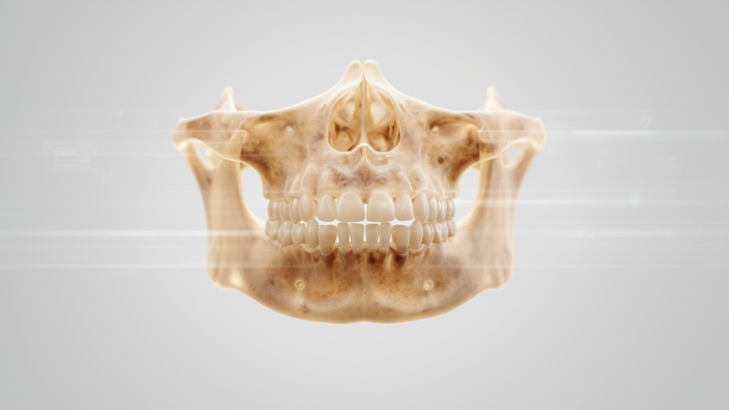
Age estimation is a critical component of human identification in forensic contexts, and a new study suggests that the maxilla, the bone that forms the central part of the face, carries measurable shape signatures of ageing. Using three-dimensional geometric morphometrics on head and neck computed tomography scans, researchers found statistically significant differences in maxillary bone shape across four adult age groups, with cross-validated classification accuracies ranging from 70.8% to 78.4%. The work, published in the Egyptian Journal of Forensic Sciences, offers a population-specific reference for Malaysian adults that could eventually support forensic identification.
The research team, led by Muhammad Faiz Mohd Fauad and colleagues, analysed 413 adult CT scans drawn from the Department of Biomedical Imaging at Pusat Perubatan Universiti Malaya. The sample comprised 208 males and 205 females, distributed across three major Malaysian ethnic groups: Malay, with 145 individuals; Chinese, with 136; and Indian, with 132. Participants ranged in age from 18 to 65 years and were sorted into four cohorts: 18 to 30, 31 to 40, 41 to 50, and 51 to 65 years. Only scans with clearly documented sex and ethnicity, complete visualisation of the maxilla, and intact craniofacial structures were included, while images showing motion artefacts, poor quality, or incomplete coverage were excluded.
To capture the geometry of the bone, the researchers digitised 45 anatomical landmarks on each specimen using the Stratovan Landmark Editor. Head and neck CT scans in standard DICOM format were imported into the software, where a three-dimensional volumetric isosurface mesh was generated by adjusting the bone density attenuation threshold. Landmarks were then placed manually on the surface mesh according to strict anatomical definitions, with the model dynamically rotated, zoomed, and viewed from anterior, lateral, and inferior planes to ensure spatial accuracy. Intra-observer and inter-observer error analyses revealed no statistically significant differences, indicating high measurement consistency, and the two independent observers were experienced anatomists working as dental and medical lecturers at the university.
The raw coordinate data were exported into MorphoJ for analysis. Generalised Procrustes Analysis first removed non-shape variation related to rotation, translation, and scaling, allowing the team to assess true shape differences. Most landmarks showed consistent deviations around the mean configuration, but greater dispersion appeared at landmarks 6 to 10 and 29 to 33, pointing to increased anatomical variability in the lateral maxillary region. Procrustes ANOVA then demonstrated a statistically significant difference in maxillary shape across the age groups, with a p-value below 0.001.
Canonical variate analysis was applied next to evaluate shape differentiation among the four cohorts, which held 104, 103, 103, and 103 samples respectively. The analysis generated three canonical variates: the first accounted for 41.11% of the total shape variation, the second for 34.26%, and the third for 24.63%. The first canonical variate showed a high degree of variation at landmarks 29 to 34 and 37 to 36, indicating changes concentrated in the left maxillary bone, including the zygomaxillary suture and the left lateral alveolar process. The second and third variates both highlighted landmarks 6 to 10 and 29 to 33, reflecting symmetric variation in the lateral bodies of the maxillary bone; in the second variate these lateral bodies were displaced outward relative to the average bone shape, while in the third they were displaced inward.
Distance measures reinforced the pattern of age-related separation. The highest Mahalanobis distance, 2.597, was observed between the youngest group, aged 18 to 30, and the eldest group, aged 51 to 65, followed by the distances between the youngest and the 41 to 50 group at 2.521, and between the 31 to 40 and 51 to 65 groups at 2.451. Procrustes distances followed the same trend, with the largest value of 0.027 again between the youngest and eldest cohorts. Permutation tests using 10,000 rounds were significant among all age groups for both distance measures. Along the first canonical axis, the youngest group was distributed toward the positive direction, the eldest toward the negative direction, and the two intermediate cohorts occupied positions between them, although the shape distribution showed substantial overlap among all groups.
Visualisation using the IDAV Landmark Editor revealed subtle morphological changes across the age spectrum. In the youngest group, the lateral margins of the bone exhibited a more pronounced curvature than in the other groups, and the alveolar process appeared broader. Surface roughness increased with age, with the eldest group displaying the most prominent irregularities, and the infraorbital foramen showed a smaller aperture in the eldest group compared with the younger cohorts. From lateral views, the alveolar process appeared smoother in the youngest group, while the variations captured from those perspectives were subtle.
Discriminant function analysis with cross-validation assessed how well maxillary shape could classify individuals into age groups, yielding accuracies of 78.4% for the youngest cohort, 71.9% for the 31 to 40 group, 70.8% for the 41 to 50 group, and 76.3% for the eldest cohort. These figures exceed those reported in a Greek skull study that applied landmarks including the maxillary region and achieved age classification accuracies of 41.2% to 62.8%. Comparable or higher accuracies have been observed in studies using maxillary sinuses of Korean patients, at 86.8% to 90.4%, and premolar teeth from micro-CT imaging, at 82%. Other geometric morphometrics studies in the Malaysian population reported age classification accuracies of 53% to 79% for mandibles and 61.5% for cervical vertebrae.
The authors note that no previous geometric morphometrics study has assessed the entire maxillary shape across age; earlier work focused on partial structures such as the maxillary sinus, dental structures, and sutures. Cone beam computed tomography studies of Chinese and Spanish populations reported reduced maxillary sinus size in older individuals, partly due to decreased pneumatization, and septa prevalence within the sinus was higher in elderly individuals. Age-related changes were also observed in maxillary dental structures, including correlations between premolar root canal position and age in Arab patients, and decreases in enamel thickness and pulp volume with age in Iraqi subpopulations. These findings highlight that both the skeletal and dental components of the maxilla undergo measurable morphological changes with ageing.
The study has limitations that shape how its results should be applied. The sample includes an equal distribution of individuals across four adult age cohorts up to 65 years but lacks representation of the elderly population over 65, a stage in which craniofacial and skeletal degenerative changes often accelerate. The researchers also did not strictly categorise or control for varying degrees of tooth loss and subsequent alveolar bone resorption, factors that can confound purely age-related skeletal modifications. Because craniofacial structures display high geographic and population-specific variation, the reference values and classification accuracies cannot be directly extrapolated to global or non-Southeast Asian populations without further comparative research. Future studies, the team says, will aim to include geriatric cohorts above 65 years and explicitly control for edentulous status to isolate pure age-related remodelling from dental-induced bone resorption.
Subject of Research: Age-related shape variation of the maxillary bone analysed with 3D geometric morphometrics in Malaysian adults
Article Title: Age related morphological variation of the maxilla: a 3-dimensional (3D) geometric morphometrics analysis
Article References: Mohd Fauad, M. F., Alias, A., Choy, K. W., Mohd Hadi, H., Chung, E., Ng, W. L., Kurniawan, A., Samshuddin, N. A. S., & Abumosameh, M. A. (2026). Age related morphological variation of the maxilla: a 3-dimensional (3D) geometric morphometrics analysis. Egyptian Journal of Forensic Sciences, 16(1), Article 78. https://doi.org/10.1186/s41935-026-00574-1
Image Credits: AI Generated
DOI: 10.1186/s41935-026-00574-1
Keywords: forensic science, age estimation, geometric morphometrics, maxilla, computed tomography, 3D landmarks, Procrustes ANOVA, discriminant function analysis, Malaysian population, craniofacial morphology, human identification, related
News Source: Beatrice Stafford. (October 30, 2026). 3D scans of the maxilla reveal how the facial skeleton ages. Scienmag.



