


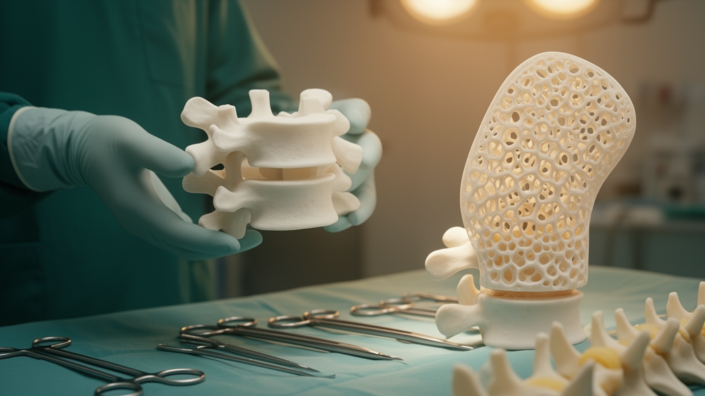
Engineers and surgeons have long been able to print anatomically accurate copies of a patient’s spine, but those replicas have mostly been visual aids: they look like bone without behaving like it. A new study from researchers at Leipzig University, published in BMC Biomedical Engineering, reports a way to close that gap. The team designed a parametrizable hexagonal lattice structure that substitutes for spongy bone inside printed vertebrae, tuned its mechanical properties through a single geometric ratio, and showed that whole printed vertebrae built this way responded to pedicle screws much like the osteoporotic human bones they copied.
The work began with real bone. The researchers obtained 43 vertebrae from six human body donors, two women and four men with a mean age of 86.8 years, spanning spinal levels from the seventh thoracic to the fifth lumbar vertebra. The donors had given written consent during their lifetimes, and the use of their post-mortem tissue was approved by the Institute of Anatomy at the University of Leipzig. All spines were stored fresh-frozen and scanned with clinical computed tomography before testing, with each vertebra later classified as normal, osteopenic or osteoporotic using dual-energy X-ray absorptiometry. Given the donors’ advanced age, the sample skewed toward reduced bone quality: two donors had normal bone density, one was osteopenic and three were osteoporotic.
From each vertebra, the team drilled a cylindrical core of spongy bone, six millimetres in diameter and twelve millimetres long, from either the left or right side of the vertebral body. Before that, a polyaxial pedicle screw had been randomly implanted on one side of each vertebra so the same bones could later serve in pull-out experiments. The cores were compressed to failure in a uniaxial testing machine, yielding three mechanical parameters: the compressive modulus, the yield stress at 0.2 percent offset, and the plateau stress averaged over the 20 to 40 percent strain range. In parallel, segmentation software measured the average volumetric Hounsfield units of the spongy bone in each vertebral hemisphere, the radiodensity value that clinical CT scanners report.
The correlation the team was hoping for emerged clearly. Mechanical properties rose with increasing Hounsfield units, and power-law curve fits captured the relationship with statistical significance. Yield stress and plateau stress fitted best, with R-squared values of 0.62 and 0.58 respectively, while the compressive modulus fitted somewhat more loosely at 0.42. The measured values themselves were low compared with published literature on vertebral cancellous bone, with compressive moduli between 3.3 and 420.9 megapascals, which the authors attribute to the geriatric age of the donors. Hounsfield units on the tested side ranged from 61 to 364, and the data separated roughly at a threshold of 200 units between denser, presumably normal vertebrae and those likely osteopenic or osteoporotic.
The second half of the study concerned the printed substitute. Rather than attempting to reproduce the intricate true architecture of trabecular bone, which would require micro-CT data and radiation doses unsuitable for patients, the researchers designed an idealised hexagonal lattice: equilateral hexagons in stacked planes connected by vertical struts. The entire structure is defined by one parameter, the ratio of strut thickness to strut length. Because stereolithography printing could not reliably reproduce struts thinner than 0.4 millimetres, that diameter was fixed, and varying the strut length produced six different porosities. The ratio also controls the lattice’s relative density, which ranged in a band closely matching the 4 to 12 percent solid content reported for vertebral cancellous bone.
Cylindrical lattice specimens were printed from two commercial photopolymer resins, Clear V4 and Tough2000 V2, and subjected to the same compression protocol as the bone cores. Across the printed samples, the mechanical properties were described excellently by the thickness-to-length ratio, again via power-law fits. The two resins bracketed the mechanical range of the human specimens well, although the printed material itself is far less stiff than real mineralised tissue, with an elastic modulus of roughly 2 to 3 gigapascals against about 17 gigapascals for femoral cortical bone. The design compensates for that gap by raising the relative lattice density. Not every combination printed cleanly: at high thickness-to-length ratios, warping during post-processing distorted some specimens, and Clear V4 samples at the densest ratio burst during testing.
With both calibration curves in hand, the parameterisation becomes a matter of arithmetic. One equation links a target mechanical property to the bone’s Hounsfield units; a second links the same property to the lattice ratio. Solving them together yields the strut geometry needed for a given vertebra, averaged across the three mechanical parameters. The team demonstrated this on three lumbar vertebrae from different donors, two osteoporotic and one of normal quality. After segmenting the outer and inner cortical contours from CT, remeshing the model and voxelising the interior with the calculated unit cells, each vertebra was printed five times in Tough2000 V2 resin, chosen after the stiffer Clear V4 proved too brittle and fractured during screw insertion in preliminary trials.
The validation test was axial screw pull-out, a standard measure of how well a pedicle screw anchors in bone and a direct proxy for spinal fixation strength. In the human specimens, maximum pull-out forces ranged from 183 to 1567 newtons and correlated significantly with local Hounsfield units, with the power-law fit reaching an R-squared of 0.83. The printed replicas landed in a revealing pattern. For the two vertebrae with poor bone quality, the replicas averaged pull-out forces about 200 newtons higher than their human originals, which the authors attribute to an over-thick cortical shell in the pedicle, likely from segmentation or post-processing error, since roughly 60 percent of pull-out resistance comes from cortical fixation. For the one vertebra with stronger bone, the replicas fell about 600 newtons short, because the resin cannot match the stiffness of real cortical bone. The abstract’s summary is therefore precise: the printed vertebrae showed pull-out forces similar to those of osteoporotic bone.
That boundary matters for how the technology should be read. The authors conclude that their approach is well suited to replicating bone with low bone quality, and they explicitly note that healthy bone or bone with pathologically elevated stiffness, as in tumours or necrosis, would require other materials, fibre reinforcements or printing technologies. The study is also an in vitro engineering validation on a small number of donor specimens, not a clinical trial, and its parameterisation rests on Hounsfield units from specific scanners at particular tube voltages, values that do not transfer directly between machines without calibration. The freehand screw insertion technique likely contributed to the variance in pull-out forces, and cortical thickness was never directly measured in either the bones or the replicas.
Within those limits, the practical promise is concrete. Patient-specific models with validated, tunable mechanical behaviour could let surgeons rehearse demanding procedures on a replica that resists drilling and screw insertion the way the actual osteoporotic vertebra will, and give biomechanical researchers a reproducible test material that reduces dependence on scarce body donors and their wide biological variation. The authors point toward future work on other lattice types, local manipulation of individual cells to mimic the heterogeneity of real trabecular bone, and extension to other anatomical regions and better bone quality. For now, the study establishes the core principle: a single geometric ratio, calibrated against routine CT numbers, is enough to make a printed vertebra that behaves, at least under a pedicle screw, like the fragile bone it copies.
Subject of Research: A parametrizable 3D-printed lattice structure that reproduces the mechanical behaviour of vertebral spongy bone in patient-specific printed vertebrae.
Article Title: Biomechanical evaluation of individual 3D-printed vertebrae
Article References: Metzner, F., Schleifenbaum, S., Heyde, C.-E., & von der Höh, N. H. (2026). Biomechanical evaluation of individual 3D-printed vertebrae. BMC Biomedical Engineering, 8(1), Article 5. https://doi.org/10.1186/s42490-026-00107-w
Image Credits: AI Generated
DOI: 10.1186/s42490-026-00107-w
Keywords: 3D printing, vertebrae, biomechanics, Hounsfield units, pedicle screws, osteoporosis, lattice structures, stereolithography, spinal surgery, cancellous bone, patient-specific models, computed tomography
News Source: Denise Maddox. (October 29, 2026). 3D-Printed Vertebrae Replicate Weak Bone Mechanics for Surgical Practice. Scienmag.



