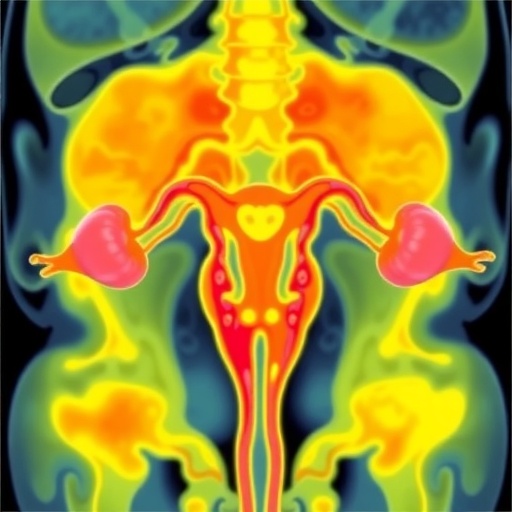
A new three-dimensional view of intact ovaries is offering scientists a more complete picture of how the ovarian reserve changes with age, challenging the idea that reproductive aging can be understood simply by counting follicles in thin tissue sections. The study, led by A. D’Angelo, D. Franco-Barranco, M. Musy and colleagues, uses whole-organ mapping to examine the spatial organization of ovarian follicles and the way that organization changes across the lifespan. Published in Nature Aging, the work places the microscopic architecture of the ovary at the center of a question with enormous consequences for reproductive medicine: why does fertility decline, and why does that decline vary so widely between individuals?
The ovarian reserve is established before birth and consists primarily of primordial follicles, each containing an immature oocyte surrounded by a small layer of support cells. Most of these follicles remain dormant for years, while a small proportion is recruited into a growth process that can eventually lead to ovulation. The reserve is therefore not merely a numerical inventory of eggs. It is a living, spatially organized population embedded within a changing tissue environment. As follicles are activated, degenerate or respond to hormonal signals, the structure of the ovary itself may influence which oocytes survive and which are lost. Capturing those relationships has been difficult because conventional histology reduces a three-dimensional organ to a series of two-dimensional slices.
The approach described by the researchers addresses that problem by reconstructing the intact ovary in three dimensions. Instead of assessing isolated sections and estimating how many follicles may exist between them, whole-organ mapping allows follicles and other anatomical features to be viewed in their original spatial context. This distinction matters technically: a follicle may appear close to another structure in one section while being separated from it in three dimensions, and the apparent density of follicles can change substantially depending on the plane of observation. Three-dimensional analysis can also reveal regional differences, clustering patterns and changes in follicle distribution that traditional sampling may overlook.
That broader view is especially important for studying aging. The ovarian reserve does not decline at a perfectly uniform rate, and ovarian aging involves more than the gradual disappearance of follicles. Follicles may be lost through a process known as atresia, in which they degenerate before ovulation. The surviving follicles may also experience changes in their surrounding cells, extracellular matrix and vascular environment. Over time, the tissue may become less capable of maintaining the conditions required for follicle dormancy, growth and oocyte quality. By mapping the reserve across intact ovaries, the study makes it possible to examine aging as a coordinated transformation of an organ rather than as a single downward curve in follicle number.
The technical significance of the work lies in connecting cellular measurements with organ-scale biology. A two-dimensional section can identify a follicle and provide information about its size, structure or developmental stage. A three-dimensional map can add information about location, neighborhood and distribution. Researchers can ask whether follicles are randomly dispersed, concentrated in particular regions or reorganized as the ovary ages. They can also compare the relative positions of follicles at different developmental stages and investigate whether aging alters the physical landscape through which follicles progress. These questions are difficult to answer with conventional microscopy alone, but they are essential for understanding how local tissue conditions shape reproductive potential.
The study’s focus on intact ovaries also has implications for the way ovarian reserve is measured in clinical practice. Current assessments often rely on indirect indicators, including circulating anti-Müllerian hormone levels, antral follicle counts observed by ultrasound and a patient’s age. These measures are useful, but each captures only part of the biology. They do not directly visualize the entire population of primordial follicles, nor do they fully describe the tissue environment in which those follicles exist. A three-dimensional atlas cannot immediately replace clinical testing, but it may reveal why commonly used markers sometimes fail to predict reproductive outcomes precisely. It may also help researchers identify structural or molecular features that could eventually improve individualized fertility assessment.
The findings are likely to resonate far beyond reproductive biology because ovarian aging is connected to broader questions about healthspan. The ovary is an endocrine organ, and changes in its function can influence hormonal signaling throughout the body. The depletion of the follicular reserve and the transition toward menopause are associated with major physiological changes, although the relationships among ovarian structure, hormones and long-term health remain complex. A detailed map of how the ovary ages could provide a foundation for studying those connections. It may also help explain why the timing of reproductive aging differs among individuals and why some ovaries appear to preserve functional characteristics longer than others.
At the same time, the work should not be interpreted as evidence that aging ovaries can currently be restored or that a three-dimensional map can predict an individual woman’s fertility. Ovarian reserve is only one component of reproductive success, and the number of remaining follicles does not necessarily indicate the genetic or developmental quality of their oocytes. Human reproductive aging is influenced by genetics, endocrine regulation, environmental exposures, disease, medical treatment and chance. The value of the research is more fundamental: it creates a framework for observing how these factors may act on an organ whose internal organization has historically been difficult to study in its entirety.
The study arrives as fertility science increasingly moves toward high-resolution, data-rich models of human biology. Combining three-dimensional imaging with molecular profiling, computational analysis and longitudinal clinical data could eventually transform the ovarian reserve from an abstract estimate into a dynamic biological system that can be measured in far greater detail. For now, the work by D’Angelo and colleagues delivers a powerful conceptual shift. The ovarian reserve is not simply a diminishing number of follicles counted under a microscope; it is a spatially structured population embedded in living tissue, and its aging may depend on how that organization changes over time. By making the intact ovary visible as a three-dimensional landscape, the researchers have opened a new route toward understanding one of biology’s most consequential and least predictable aging processes.
Subject of Research: Three-dimensional organization and age-related dynamics of the ovarian reserve.
Article Title: Three-dimensional mapping of intact ovaries reveals the aging dynamics of the ovarian reserve.
Article References: D’Angelo, A., Franco-Barranco, D., Musy, M. et al. “Three-dimensional mapping of intact ovaries reveals the aging dynamics of the ovarian reserve.” Nature Aging 6, 1580–1591 (2026). https://doi.org/10.1038/s43587-026-01178-z
Image Credits: AI Generated
DOI: https://doi.org/10.1038/s43587-026-01178-z
Keywords: ovarian reserve, ovarian aging, fertility, reproductive biology, three-dimensional imaging, follicles, oocytes, menopause, tissue mapping, aging research
Tags: 3D ovarian mappingfertility decline variationfollicle activation and degenerationmicroscopic ovarian structureovarian lifespan and functionovarian reserve agingovarian tissue architectureprimordial follicle dynamicsreproductive aging mechanismsreproductive medicine implicationsspatial organization of ovarian follicleswhole-organ ovary analysis