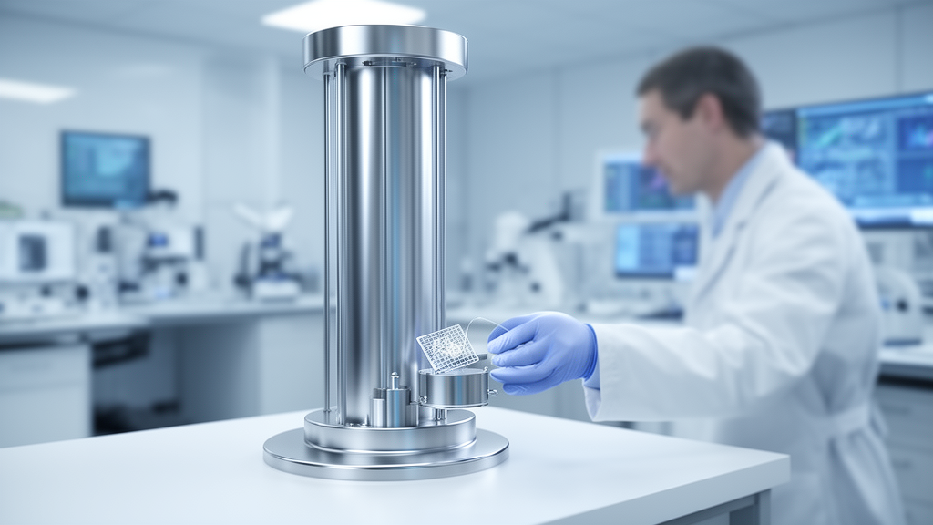
The wiring of a brain can now be traced synapse by synapse, but only a handful of laboratories on Earth possess the instruments and computing muscle to do it. That exclusivity is about to end. The University of Vermont has been named the national coordinating hub for a five-year, $19 million project funded by the National Science Foundation to design and build the National Connectomics and Ultrastructure Volume Observatory, or NCUVO, a facility intended to bring ultra-high-resolution, three-dimensional biological imaging to researchers across the United States. If the design succeeds, questions once confined to elite imaging centers, from how learning physically rewires memory circuits to how a single-celled parasite breaches the blood-brain barrier, could be pursued by scientists at any institution in the country.
The project is led by neuroscientist Davi Bock of UVM’s Robert Larner, M.D. College of Medicine, with co-principal investigators Doug Taatjes, director of UVM’s Microscopy Imaging Center, and Mark Ellisman at the University of California San Diego. Together the partners will integrate three complementary capabilities: state-of-the-art high-throughput electron microscopy at the Larner College of Medicine, advanced sample preparation and multimodal imaging expertise at UC San Diego’s National Center for Microscopy and Imaging Research, and large-scale data management at the San Diego Supercomputer Center. The ambition is to design a complete pipeline that carries a preserved biological sample all the way to analyzed, explorable scientific data.
Bock has spent two decades helping to build the science of connectomics, the mapping of nervous systems down to individual neurons and the synapses that link them. His laboratory acquired the electron microscopy images that an international collaboration used to produce the first complete wiring map of an adult fruit fly brain, a landmark achievement published in Nature in 2024. That accomplishment demanded enormous gains in imaging speed and computational power, because the hard problem has never been magnification alone. Electron microscopes already resolve astonishingly small structures; the challenge lies in assembling and aligning millions of images to follow the branching paths of neurons through substantial volumes of tissue, and in converting those image stacks into usable maps.
Having built that capacity, Bock argues the moment has come to widen both its reach and its range. “We built a searchlight to look at one type of problem, and now, with a purpose-built facility for advanced work like this, we will use it to look at many, many different problems,” Bock says. “This new national center will be globally unique in pointing high throughput volume microscopes at the full spectrum of biology. Right now, that doesn’t exist anywhere else.” The observatory’s name is deliberate: just as an astronomical observatory gives many researchers shared access to the heavens, NCUVO will give biologists a shared window onto structures far too small for ordinary light microscopes, the fine details known as ultrastructure that fill the interiors of cells and the spaces between them.
The technical workflow will unfold across three institutions. Tissue preserved in resin will be shaved into thousands of ultrathin slices, each captured at nanometer-scale resolution by a high-throughput electron microscope housed in UVM’s Firestone Medical Research Building. Computers at the San Diego Supercomputer Center will then assemble the slices into coherent three-dimensional volumes. Artificial intelligence will shoulder much of the interpretive burden, identifying cell boundaries, tracing individual neurons through dense tissue, and distinguishing organelles and other features within cells. Researchers will retain tools to verify the automated reconstructions, label structures of interest, and interrogate the data with their own questions. At the San Diego microscopy center, selected regions can be re-imaged at even higher resolution, connecting tiny details to the larger anatomical context mapped in Vermont.
The data volumes will be staggering, measured in petabytes, or millions of gigabytes, per dataset. UVM’s Vermont Advanced Computing Center will support storage and computation, while Ilkay Altintas, chief data science officer at the San Diego Supercomputer Center and principal investigator of the NSF-funded National Data Platform, will guide data curation, drawing on her team’s expertise in organizing, processing and sharing massive scientific datasets and linking the observatory to national cyberinfrastructure. “You can’t just drop a petabyte of data on someone and say mission accomplished,” Bock says. “You have to give them tools to analyze the data.” Ellisman frames the design philosophy the same way: the goal is to transform the latest sample preparation, multimodal imaging and computational technologies into a shared national capability rather than a specialty reserved for a few, empowering scientists to compare many samples at levels of detail ranging from whole tissues down to individual synapses.
Four pilot projects will stress-test the pipeline against different organisms, sample sizes and scientific demands. The first will examine how connections in the mouse hippocampus, a brain region essential for memory, change after learning, in collaboration with Anton Maximov’s laboratory at Scripps Research and Ellisman’s team at UC San Diego; researchers will trace circuits involving neurons flagged as active during a learning task. The second will compare retinal circuitry across species to probe how visual systems adapt to different environments, pairing David Berson’s laboratory at Brown University with Tom Baden’s laboratory at the University of Sussex. Bock points to sharks navigating murky versus clear water as the kind of comparative question these cross-species maps could finally answer. The work will generate datasets large enough, and samples numerous enough, for statistical comparison of how brain structures differ with learning, development or disease.
The third and fourth pilots reach beyond classic neuroscience entirely. Parasitologist Bruno Martorelli Di Genova’s laboratory at UVM will join Christopher Moore’s laboratory at Brown University to study how the parasite Toxoplasma gondii crosses the blood-brain barrier to cause toxoplasmosis, a disease spread through contaminated food and cat litter. “This is an amazing organism because it causes rodents to lose their fear of cat urine,” Bock explains. “It changes the behavior of cat prey to be less frightened of cats. And there’s some evidence that infected humans also are less risk averse.” The fourth pilot will image protists, single-celled organisms with surprisingly elaborate internal architecture, in partnership with Omaya Dudin’s laboratory at the University of Geneva and Gautam Dey’s laboratory at the European Molecular Biology Laboratory in Heidelberg, both recipients of the 2026 EMBO Gold Medal for their work in evolutionary cell biology.
Education is built into the observatory’s mission as well. UVM neuroscience assistant professor Molly Stanley will coordinate a collaboration with Connectomes for Undergraduate Neuroscience Education and Learning, whose leadership includes Andrew Bellemer at Appalachian State University, Kenneth Colodner at Mount Holyoke College and Divya Sitaraman at California State University, East Bay, to develop undergraduate teaching materials from the pilot datasets. The project also plans virtual-reality materials for high school classrooms built on the syGlass platform, along with training opportunities for UVM biomedical engineering master’s students, ensuring that the resource cultivates the next generation of researchers even before the full facility exists.
The five-year effort will deliver a tested design, pilot datasets and an implementation plan for a subsequent national facility, with approximately $15.68 million supporting Bock’s work at UVM and $3.32 million funding UC San Diego’s contributions, including the supercomputer center. The research is fundamentally curiosity-driven basic biology, but the team expects it could illuminate the structural changes that accompany neurodegenerative disease and neural repair, and even inform new approaches to artificial intelligence. “You can’t predict what we’ll find. And that’s the point,” Bock says, invoking Antony van Leeuwenhoek’s first glimpses of microscopic life, observations that seemed abstract at the time but ultimately led to the germ theory of disease. “Anything that you can think of, any tissue, any species, any organism we can look at, we should,” he says. “That’s democratization, broadening the spectrum of questions that can be asked and answered by our national research community.”
Subject of Research: A National Science Foundation-funded connectomics and ultrastructure imaging observatory
Article Title: University of Vermont launches $19 million national research center to unlock secrets of neurobiology
Article References: University of Vermont launches $19 million national research center to unlock secrets of neurobiology. (n.d.). Original publication
Image Credits: AI Generated
DOI: Not provided
Keywords: connectomics, electron microscopy, neuroscience, National Science Foundation, University of Vermont, three-dimensional imaging, artificial intelligence, ultrastructure, petabyte data, Toxoplasma gondii, brain mapping, scientific observatory
News Source: Cassandra Pierce. (October 18, 2026). $19 Million NSF Observatory Aims to Map Life’s Smallest Structures for Scientists Nationwide. Scienmag.