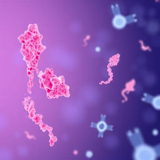
In the relentless pursuit to unravel the mysteries behind Parkinson’s disease, a groundbreaking study has emerged that shines new light on the enigmatic role of α-synuclein fibrils. Researchers led by Ohira, Sawamura, Kawano, and colleagues have published compelling evidence demonstrating how structural variations within α-synuclein fibrils profoundly influence their seeding activity — a key driver of neurodegenerative progression in Parkinson’s disease models. The implications of this discovery underscore not only the heterogeneity in pathological α-synuclein assemblies but also herald a transformative shift toward precision-targeted therapeutic strategies.
The recent investigation meticulously dissected this complexity by employing a combination of biochemical assays, electron microscopy, and in vivo experimental models. Their approach rested on isolating distinct α-synuclein fibrillar conformations and characterizing their seeding capacity across cellular and animal paradigms that recapitulate Parkinsonian neurodegeneration. The juxtaposition of structural biochemistry with functional outcomes provided unprecedented insight into how minute alterations at the fibril level translate into large-scale pathogenic cascades.
.adsslot_a6zb57Mf9i{ width:728px !important; height:90px !important; }
@media (max-width:1199px) { .adsslot_a6zb57Mf9i{ width:468px !important; height:60px !important; } }
@media (max-width:767px) { .adsslot_a6zb57Mf9i{ width:320px !important; height:50px !important; } }
ADVERTISEMENT
One of the pivotal revelations was the demonstration that α-synuclein fibrils are not a monolithic species but encompass a spectrum of polymorphs with differential seeding efficiencies. These conformers vary in their secondary and tertiary structures, leading to distinct kinetic profiles of aggregation and divergent neurotoxic potential. Notably, certain conformers exhibited heightened propensity to propagate pathology, accelerating neurodegeneration and motor dysfunction in experimental hosts.
The morphologies delineated by high-resolution electron microscopy revealed fibrillar architectures ranging from tightly packed twisted ribbons to loosely associated protofilament bundles. This structural plasticity is likely driven by variations in fibril core regions, post-translational modifications, and interactions with cellular cofactors. The study attentively correlates these ultrastructural nuances with biophysical properties such as stability, fragmentation tendency, and affinity for cellular membranes — all factors that mediate seeding activity and neuroinvasion.
In vivo validation further solidified the conceptual framework by demonstrating that inoculation with specific α-synuclein polymorphs induced discrete patterns of pathology spread within rodent models. This seeding selectivity appeared to influence not only the rate but also the anatomical distribution of neurodegeneration. For instance, fibrils with enhanced seeding activity preferentially targeted nigrostriatal pathways, replicating the stereotypical lesion pattern of human Parkinsonism with remarkable fidelity.
The pathogenic variability observed suggests that Parkinson’s disease, traditionally viewed as a uniform clinical entity, might instead represent a spectrum of synucleinopathies driven by distinct conformational strains. This notion aligns with emerging paradigms in prion biology where strain diversity dictates disease phenotype and progression dynamics. Translating these insights into clinical contexts could pave the way for diagnostic tools capable of detecting specific α-synuclein strains in patient-derived biofluids, enabling earlier and more precise interventions.
From a therapeutic vantage point, the findings compel a reevaluation of anti-α-synuclein strategies. Current approaches largely target monomeric or aggregated forms indiscriminately, potentially overlooking the nuanced pathogenic hierarchy imposed by fibril polymorphism. Tailoring inhibitors or immunotherapies to the structural motifs characteristic of highly potent fibril conformers holds promise for enhanced efficacy and disease-modifying outcomes.
Furthermore, the study underscores the importance of seeding assays as predictive platforms for assessing disease trajectory and therapeutic response. By quantifying seeding activity and correlating it with clinical manifestations, researchers and clinicians may establish robust biomarkers to monitor progression and treatment efficacy in real time, a critical advancement in neurodegenerative disease management.
The research also provides a framework for investigating environmental and genetic factors influencing fibril diversification. Variations in α-synuclein sequence, cellular milieu, and cofactor presence could skew the folding landscape toward specific strains, accounting for interindividual heterogeneity in Parkinson’s pathology. Deciphering these modulators would enrich our understanding of disease etiology and identify novel targets for intervention.
Importantly, the elucidation of fibril strain-specific neurotoxicity invites exploration into synaptic and axonal vulnerabilities. Given that different fibril conformers appear to preferentially impact designated neuronal circuits, dissecting the mechanistic underpinnings of such selectivity could reveal new molecular nodes amenable to therapeutic modulation, potentially arresting or reversing neuronal loss.
This study’s meticulous integration of structural biology, neurobiology, and translational modeling epitomizes the multidisciplinary approach necessary to confront complex proteinopathies. It also opens avenues for investigating analogous mechanisms in related disorders such as multiple system atrophy and dementia with Lewy bodies, where α-synuclein aggregation also plays a central role.
As the frontier of Parkinson’s research expands, the detailed characterization of α-synuclein fibril polymorphism will undoubtedly be a cornerstone in developing next-generation diagnostics and targeted therapeutics. Although challenges remain in fully mapping the conformational landscape and translating findings into clinical applications, the current work marks a decisive milestone in the quest to decode one of neurodegeneration’s most formidable enigmas.
By redefining α-synuclein pathology through the prism of fibril structure and seeding dynamics, this study sets the stage for a paradigm shift. The era of “one-size-fits-all” approaches to Parkinson’s disease is giving way to precision molecular stratification, enabling interventions that can selectively neutralize the most pernicious α-synuclein species driving disease progression.
Ultimately, these findings illuminate the intricate relationship between protein structure, seeding activity, and neurodegeneration, guiding a roadmap toward more effective management of Parkinson’s disease. A deeper understanding of α-synuclein’s structural diversity not only enriches fundamental neuroscience but also inspires hope for millions affected by this relentless disorder, underscoring the transformative potential of molecularly informed medicine.
Subject of Research: Impact of α-synuclein fibril structure on seeding activity in experimental models of Parkinson’s disease.
Article Title: Impact of α-synuclein fibril structure on seeding activity in experimental models of Parkinson’s disease.
Article References:
Ohira, J., Sawamura, M., Kawano, K. et al. Impact of α-synuclein fibril structure on seeding activity in experimental models of Parkinson’s disease. npj Parkinsons Dis. 11, 224 (2025). https://doi.org/10.1038/s41531-025-01080-2
Image Credits: AI Generated
Tags: anti-aggregation treatments for Parkinson’sbiochemical assays in Parkinson’s studieselectron microscopy in fibril analysisLewy bodies formationmolecular diversity of α-synucleinneurodegenerative progressionParkinson’s disease researchprecision-targeted therapeutic strategiesprion-like mechanisms in neurodegenerationseeding activity of α-synucleinstructural variations in fibrilsα-synuclein fibril structure


