The research, conducted at the Laboratory of Fish Ecophysiology at Bangladesh Agricultural University, investigated how these antioxidants protect against mercuric chloride-induced impairments in growth performance, blood parameters, and tissue integrity. The findings suggest that both supplements offer complementary protective mechanisms, restoring physiological homeostasis in this globally significant aquaculture species.
Mercury is a ubiquitous environmental contaminant that undergoes complex biogeochemical transformations in aquatic systems, eventually concentrating within food webs. In fish, mercury exposure can produce multi-system toxicity, manifesting as reduced growth, altered metabolism, impaired osmoregulation, and marked histological damage to gill, liver, and kidney tissues. The toxic potency of mercury is mediated by its chemical forms and bioavailability, with inorganic mercury potentially being methylated by sediment microorganisms into methylmercury, a species that bioaccumulates and poses significant risks to consumers.
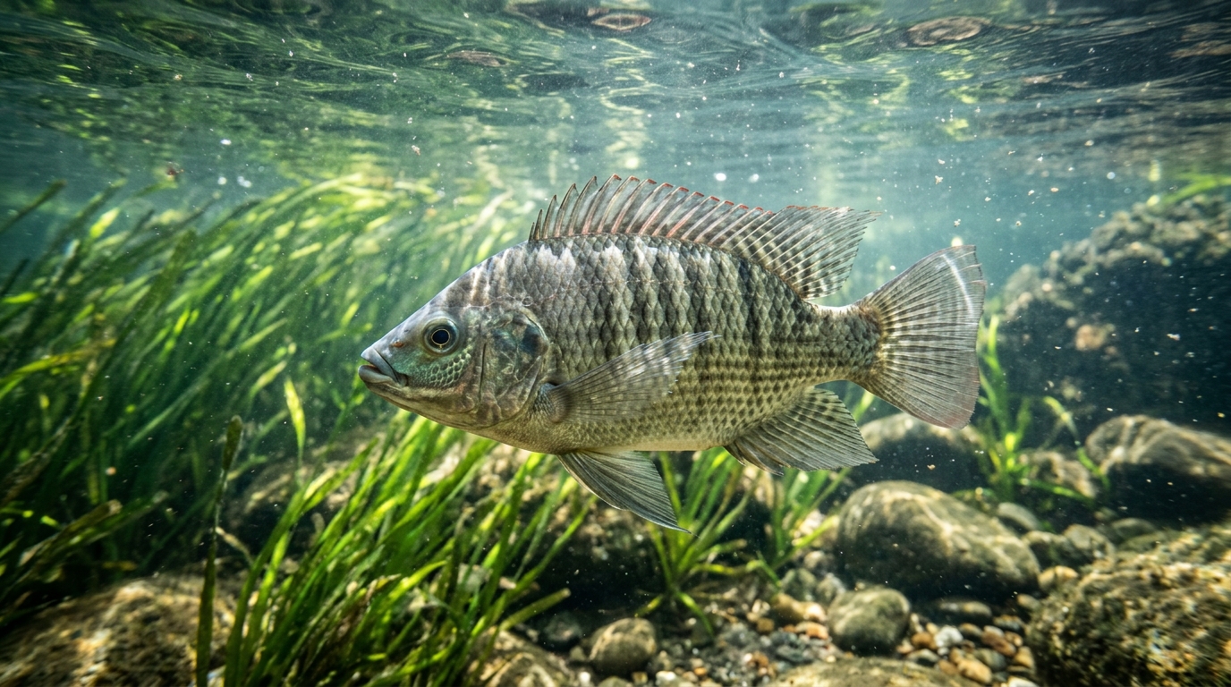
The study aimed to test the hypothesis that dietary vitamin E and selenium nanoparticles confer protection against sublethal mercuric chloride exposure. The researchers selected Nile tilapia (Oreochromis niloticus) as the model organism due to its immense aquaculture significance and vulnerability to environmental contaminants. The experimental design included four treatments, each with three replications: a control group, a mercury-exposed group, a group supplemented with mercury and vitamin E, and a group supplemented with mercury and selenium nanoparticles. The mercury concentration was set at 0.03 mg/L, corresponding to one-tenth of the 96-hour LC50 value for Nile tilapia, to induce sublethal but biologically significant stress.
Two hundred and forty healthy Nile tilapia were acclimated for 15 days before the 42-day exposure trial. The fish were housed in 12 glass aquaria, with 20 fingerlings per tank. These doses were selected from previous studies that showed such supplementation improved growth performance and overall physiology in Nile tilapia. The fish were fed twice daily, and water quality parameters including temperature, pH, and ammonia were monitored throughout the experiment.
Results indicated that mercury exposure significantly reduced final body weight, weight gain, and specific growth rate while increasing the feed conversion ratio compared to the control group. The mercury-exposed group also experienced significantly lower survival rates. However, supplementation with either vitamin E or selenium nanoparticles significantly enhanced these growth parameters compared to the mercury-only group.
Metabolic markers also reflected the stress induced by mercury exposure. The mercury group showed significantly increased levels of blood haemoglobin and glucose, characteristic of a systemic stress response involving the mobilisation of energy reserves and impaired oxygen transport capacity. Both vitamin E and selenium nanoparticle supplementation significantly reduced these increments, bringing the values closer to those of the control group. This restoration suggests that the supplements helped mitigate systemic stress and improve physiological homeostasis by restoring redox conditions and stabilising erythrocyte membranes.
Analysis of erythrocyte cellular and nuclear morphology revealed that mercury exposure increased the frequency of abnormalities such as twin-shaped cells, fusion cells, teardrop-type cells, and elongated cells. Vitamin E supplementation significantly restored the damage caused by mercury in several of these categories, although some abnormalities remained significantly higher than in the control group. Selenium nanoparticles also showed protective effects, particularly in reducing the frequency of nuclear abnormalities such as notched nuclei and nuclear buds. These findings indicate that both supplements play a mechanistic role in preserving nuclear integrity and limiting peroxidative damage to membrane lipids.
Histopathological assessment of gills revealed typical mercury-induced lesions, including congestion in primary lamellae, clubbing in secondary lamellae, and hypertrophy and hyperplasia in both primary and secondary lamellae. The severity of these lesions was most pronounced in the mercury-exposed group. Vitamin E supplementation resulted in mild changes in both primary and secondary lamellae, while selenium nanoparticles completely cured the clubbing in secondary lamellae. Both supplements restored hypertrophy and hyperplasia to null or mild levels, demonstrating their effectiveness in mitigating gill tissue damage.
In the intestine, mercury exposure significantly reduced villus length, wall thickness, muscle thickness, crypt depth, and goblet cell abundance, while increasing lamina propria width. Vitamin E restored villus length, wall thickness, and muscle thickness to values not significantly different from the control group. Selenium nanoparticles significantly improved crypt depth and recovered the structural thickness of the wall and muscularis, although villus length remained similar to the mercury group. Both supplements normalised lamina propria width and restored goblet cell abundance, indicating their role in attenuating mercury-induced intestinal damage and preserving mucosal barrier function.
Principal component analysis of the data showed that treatment groups clustered separately, with the mercury-treated group correlating with stress-related traits and the control group aligning with growth and condition variables. Both supplemented groups shifted closer to the control cluster, with selenium nanoparticles showing greater restoration of growth traits. These patterns suggest that selenium nanoparticle co-treatment provides more effective mitigation of mercury-induced physiological disturbances. The study acknowledges limitations, including the lack of analytical verification of chemical concentrations in water and the absence of mercury measurement in fish tissue for uptake and elimination. Future studies are planned to include toxicokinetic assessments for a better understanding of metal distribution in fish bodies.
The formulation of the experimental feeds and the management of aquarium conditions followed precise chemical and husbandry protocols. To generate the sublethal mercury exposure medium, researchers dissolved 1.35 grams of mercuric chloride in 1000 milliliters of distilled water, producing a 1 milligram per milliliter stock solution that was maintained in a light-resistant container to halt photodegradation. The selenium diet was constructed by combining 6.57 milligrams of sodium selenite per kilogram of commercial feed, utilizing rice gruel at one-tenth of the feed mass as an adhesive binder. In parallel, the vitamin E treatment incorporated 106 microliters of DL-alpha-tocopherol acetate per kilogram of commercial diet, using corn oil at one-tenth of the total feed weight to facilitate uniform coating before the pellets were air-dried in a temperate environment. Throughout the 42-day trial, the juvenile fish held in 100-liter glass aquaria were allowed one hour following each morning and evening feeding to ingest their rations, after which uneaten feed particles and metabolic wastes were siphoned out to preserve water hygiene.
Standardized biological collection and staining techniques governed the tissue and cellular evaluations conducted at trial completion. Prior to harvesting, feeding was withheld for 24 hours, and fish were sedated with clove oil at 5 milligrams per liter to mitigate physical trauma and involuntary movement. Blood aliquots of 5 microliters were gathered from the caudal peduncle using micropipettes and mixed with equal volumes of anticoagulant EDTA-phosphate buffered saline at a neutral pH between 6.8 and 7.0. Cytological smears were prepared on glass slides, air-dried for 15 minutes, fixed in pure ethyl alcohol, and stained with 5 percent Giemsa solution for 10 minutes. Under 40-times optical magnification, technicians evaluated roughly 500 red blood cells across five viewing fields per slide, recording the prevalence of anomalies including karyopyknosis alongside erythrocyte distortions. For microscopic organ evaluations, excised gill arches and intestinal sections were cleared of luminal debris, fixed in Bouin’s fluid for 24 hours, preserved in 70 percent ethanol, and dehydrated through an ascending alcohol series before paraffin embedding. Microtome sections cut at 5 micrometers were subjected to Mayer’s hematoxylin and eosin staining, with complementary alcian blue staining applied specifically to enumerate intestinal goblet cells across 20 to 40 slides per group.
Detailed numerical comparisons across the rearing regimes confirmed that dietary additives offset growth depression and tissue thinning. While all groups started with a baseline weight of 10.82 grams, the final weight in the mercury group dropped to 24.96 grams, yielding a weight gain of only 14.14 grams. In comparison, fish supplemented with selenium nanoparticles attained a final weight of 34.44 grams and a gain of 23.62 grams, while vitamin E recipients achieved 32.90 grams and 22.08 grams. Daily specific growth rates fell to 0.85 percent under mercury exposure relative to 1.06 percent in controls, but climbed to 1.14 percent with vitamin E and 1.18 percent with selenium nanoparticles. Concurrently, feed conversion efficiency worsened to 1.73 in the mercury cohort compared with 1.18 in controls, whereas the vitamin E and selenium groups recorded favorable ratios of 1.02 and 1.00. Intestinal morphometry mirrored this physiological divergence. Unsupplemented mercury exposure collapsed intestinal wall thickness to 6.74 micrometers and muscularis thickness to 14.31 micrometers, compared to control values of 16.73 and 23.38 micrometers. Vitamin E restored these dimensions to 17.05 and 23.77 micrometers, while selenium nanoparticles sustained them at 15.79 and 24.06 micrometers, alongside an expansion in crypt depth reaching 79.30 micrometers.
The protective responses reflect complementary biochemical actions between the trace element and the lipophilic vitamin. Mercury damages cellular components by depleting reduced glutathione, disrupting redox homeostasis, and suppressing vital antioxidant selenoenzymes like glutathione peroxidase. By binding directly to mercury, selenium forms inert complexes that reduce the bioavailability and systemic toxicity of the metal, while simultaneously bolstering selenoprotein synthesis to eliminate peroxides. In parallel, vitamin E functions within cellular membranes to halt lipid peroxidation chain reactions, preserving enterocyte membrane fluidity, nutrient absorptive capacity, and structural tissue barriers.
Subject of Research: Protective Effects of Vitamin E and Selenium Nanoparticles Against Mercury Induced Growth, Blood and Histopathological Changes in Tilapia, Oreochromis niloticus
Article Title: Protective Effects of Vitamin E and Selenium Nanoparticles Against Mercury Induced Growth, Blood and Histopathological Changes in Tilapia, Oreochromis niloticus
Article References: Ananna, M. A., Dash, A., Hossain, A. A., Amin, M. R., Shahjahan, M., & Al‐Emran, M. (2026). Protective Effects of Vitamin E and Selenium Nanoparticles Against Mercury Induced Growth, Blood and Histopathological Changes in Tilapia, Oreochromis niloticus. Veterinary Medicine and Science, 12(6), Article e71305. https://doi.org/10.1002/vms3.71305
Image Credits: AI Generated
DOI: 10.1002/vms3.71305
Keywords: Protective, Effects, Vitamin, Selenium, Nanoparticles, Against, Mercury, Induced, Growth, Blood, Histopathological, Changes
News Source: Drew Townsend. (November 7, 2026). Vitamin E and Selenium Nanoparticles Mitigate Mercury Toxicity in Tilapia. Scienmag.