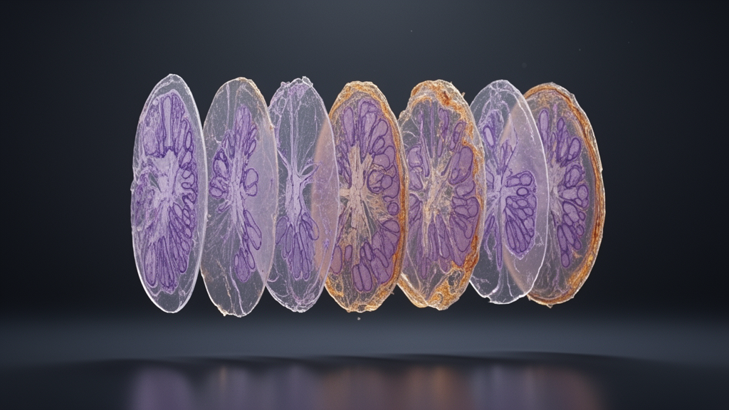
In standard clinical histopathology, macroscopic surgical resections are regularly sliced and partitioned into separate microscopic cassettes for processing and analysis, a grossing procedure that inadvertently disrupts the spatial continuity across contiguous tissue regions. When pathologists inspect individual glass slides or digitized whole-slide images under high magnification, they examine isolated tissue fragments that lack direct physical coordinates relating them back to their neighboring blocks. This fragmentation proves acutely problematic in malignant disease management and surgical oncology, particularly during complex head and neck resections, where determining continuous tumor extent and confirming negative margin boundaries directly affect patient prognosis. Because conventional macroscopic grossing introduces tears, stretching, irregular boundaries, and missing interfaces without establishing ground-truth positional tracking, reconstructive spatial correlation has traditionally persisted solely as an imprecise mental model shared between operating surgeons and evaluating pathologists through descriptive text reports.
To overcome this long-standing clinical hurdle, researchers Christoph Blattgerste, Tanzina Ferdous, Ayk Jessen, Maximilian Legnar, Karl Rohr, Claudia Scher, Jürgen Hesser, and Cleo-Aron Weis engineered HiViTAlign, an open-source deep learning framework designed to reconstruct complete histological cross-sections from fragmented tissue slices. Unlike established whole-slide image registration suites that depend on overlapping physical structures, contiguous tissue layers, or serial sections extracted from the same physical block, HiViTAlign treats histological reassembly as a single-solution pictorial puzzle composed of irregular, non-overlapping planar pieces. Operating without reference photographs or prior spatial blueprints, the modular framework unites computer vision with discrete optimization, formulating the reassembly workflow into four sequential operational phases: digital slide preprocessing and tissue foreground masking, pairwise boundary and texture registration via a hierarchical vision transformer, quantitative pairwise adjacency disparity scoring, and global puzzle configuration using discrete graph clustering algorithms.
The procedural pipeline begins by standardizing incoming whole-slide images through downsampled thumbnails generated at a uniform zoom factor using the OpenSlide library. Because digital microscopy slides inherently present extensive empty white backdrops that can distort registration algorithms, automated tissue segmentation isolates viable specimen contours against a suppressed black background. The investigators evaluated basic thresholding via Otsu’s method alongside an adaptive Canny edge detection pipeline operating on grayscale conversions. Comparative analysis demonstrated that converting image data into the LAB color space provided superior masking precision on lightly stained hematoxylin and eosin sections containing delicate adipose regions, effectively preserving faint cellular edges while suppressing unstained glass areas. Once masked and centered, the individual tissue fragments enter the pairwise alignment stage, where their mutual orientations and positional offsets must be calculated based purely on morphological and chromatic coherence rather than intersecting structural pixels.
To calculate the spatial alignment between unjoined fragment pairs, the research team adapted the Coarse-to-Fine Vision Transformer, known as C2FViT, an architecture originally designed for three-dimensional medical scan registration. While classical feature-based detection algorithms such as SIFT or SURF search for redundant feature correspondences that do not exist across separated non-overlapping tissue margins, the vision transformer leverages multi-head self-attention mechanisms to register long-range textural relationships and global contextual patterns. The adapted network resolves a multi-resolution regression task across fixed and moving two-dimensional RGB fragment tiles, progressively predicting continuous translational shifts along horizontal and vertical axes to achieve optimal edge alignment. Although the mathematical formulation accommodated rotational angles and translational vectors, experimental optimization indicated that while translation parameters converged with high fidelity, rotational and reflective parameters could not be estimated with sufficient reliability, leading the authors to train the model to predict rigid translational shifts.
Supervised training of complex vision transformers typically requires tens of thousands of precisely annotated ground-truth image pairs, a resource entirely absent from digital pathology archives. To solve this data scarcity, the team constructed a scalable synthetic puzzle generator that converts arbitrary whole-slide images into realistic multi-piece puzzles with known translational coordinates. The generator overlays geometric polygons derived from German postal-code boundary networks downloaded from OpenStreetMaps onto masked whole-slide image thumbnails. Because administrative postal boundaries feature complex, winding, and jagged perimeters, cropping tissue slides along these paths closely mimics natural mechanical tears, microtome fracturing, and cutting artifacts encountered during surgical grossing. The pipeline automatically labels, centers, and exports each extracted irregular fragment, creating vast synthetic training benchmarks featuring fully verifiable translational ground-truth offsets without necessitating arduous manual annotations by medical specialists.
Using this synthetic generator, the investigators trained the adjusted C2FViT network across three diverse datasets to instill broad morphological generalization and staining resilience. The training corpus comprised 32,560 irregular puzzles generated from 6,512 tissue microarray patches across 230 whole-slide images at a resolution of 1.84 micrometers per pixel, accompanied by 44,384 regular two-by-two square tile puzzles cropped with slight overlapping margins between 0 and 10 pixels to promote fine cellular texture matching. To incorporate real-world biological complexity, the team integrated a third dataset consisting of 21,620 puzzles derived from 192 ex vivo porcine organ whole-slide images encompassing three different organ structures, scanned at a resolution of 14.7 micrometers per pixel using tissue harvested under local animal research ethics approvals. The model was trained for 256 epochs on a high-performance cloud server equipped with an NVIDIA RTX A6000 graphics processing unit, an AMD EPYC 7502 8-core central processor, and 128 gigabytes of system memory, consuming approximately 200 seconds per training epoch.
Following pairwise transformation prediction, HiViTAlign evaluates candidate fragment matches using a custom objective similarity metric combining geometric boundary contact with chromatic compatibility. Because standard ground-truth assessment metrics like the Structural Similarity Index Measure or Peak Signal-to-Noise Ratio cannot be applied to unreferenced surgical slides, the formulated function balances the proportion of perimeter coordinates falling within an adaptive contact distance against any overlapping tissue area, multiplied by the inverse color difference across adjacent boundary points. The mathematical reciprocal of this score defines an edge disparity cost, which populates an exhaustive pairwise disparity matrix. To determine the global arrangement of all fragments without incurring the prohibitive computational overhead of integer programming, the framework implements discrete combinatorial clustering algorithms, evaluating agglomerative hierarchical clustering, stochastic simulated annealing with an adaptive cooling schedule, and a greedy minimal spanning tree utilizing Kruskal’s algorithm and path-compressed union-find data structures.
Quantitative performance evaluations over 500 test puzzles per dataset demonstrated substantial differences across assembly strategies and input characteristics. In pairwise registration tests, the vision transformer outperformed classical gradient-based optimization by a factor of 2.65 in translation mean absolute error, achieving structural normalized cross-correlation values between 0.96 and 0.97. When comparing global assembly algorithms on synthetic irregular puzzles averaging 4.74 fragments, the minimal spanning tree strategy demonstrated clear superiority over agglomerative clustering and simulated annealing, achieving a 9.0 percent reduction in puzzle-level translation error, a 28 percent reduction in mean squared error, and tighter cycle-consistency error spreads. Furthermore, executing the minimal spanning tree algorithm required only 2.78 seconds per puzzle on mid-tier hardware configured with an NVIDIA Quadro M6000 GPU and an Intel Core i7-6800K processor, proving notably faster than simulated annealing at 3.68 seconds and agglomerative clustering at 3.09 seconds, underscoring its suitability for hospital IT infrastructure.
To evaluate practical real-world utility, the full HiViTAlign pipeline was tested on 500 ex vivo porcine organ puzzles averaging 3.33 pieces per image. The complete automated framework achieved an overall puzzle accuracy of 45 percent with a standard deviation of 22 percent, registering an average translation uncertainty of 4.6 millimeters across the 1024-by-1024 pixel specimen fields, which corresponded to approximately 3.3 millimeters per coordinate axis. In a direct operational benchmark involving 48 puzzles, automated reassembly executed over 30 times faster than manual human reassembly of pre-segmented fragments. However, manual alignment retained superior precision, exhibiting 20.7 percent lower mean squared error and 5.6 percent higher normalized cross-correlation. A key vulnerability identified across experiments was compounding error propagation within the minimal spanning tree: while puzzles comprising three fragments attained a 97 percent assembly success rate, accuracy dropped below 70 percent for complex puzzles containing more than six pieces, as slight initial pairwise registration shifts compounded unchecked across sequential branch traversals.
The study authors emphasized that while HiViTAlign represents the first automated pipeline capable of reassembling disconnected, irregular, and unreferenced histopathology slices, its current spatial margin of error precludes immediate standalone diagnostic deployment for critical surgical decisions. Instead, the open-source pipeline establishes a functional proof-of-concept that can immediately assist pathologists through semi-automated workflows, rapidly proposing top-ranked fragment adjacencies and candidate orientations for manual validation. Moving forward, the researchers highlighted the necessity of integrating closed-loop pose-graph optimizations to eliminate compounding branch errors, incorporating affine transformation modules to model physical specimen distortion, and implementing physics-informed neural networks to constrain fragment boundaries according to realistic biochemical tissue compliance, ultimately establishing foundational digital bridges connecting two-dimensional histology slices to comprehensive three-dimensional surgical and radiological models.
Subject of Research: Automated histopathological tissue fragment reassembly using a vision transformer alignment pipeline
Article Title: Post-operative tissue fragment puzzling using histopathological vision transformer alignment HiViTAlign
Article References: Blattgerste, C., Ferdous, T., Jessen, A., Legnar, M., Rohr, K., Scher, C., Hesser, J., & Weis, C.-A. (2026). Post-operative tissue fragment puzzling using histopathological vision transformer alignment HiViTAlign. BMC Digital Health, 4(1), Article 49. https://doi.org/10.1186/s44247-026-00277-6
Image Credits: AI Generated
DOI: 10.1186/s44247-026-00277-6
Keywords: Digital Pathology, Vision Transformer, Image Registration, HiViTAlign, Histopathology, Surgical Margins, Whole Slide Images, Minimum Spanning Tree, Post-operative, tissue, fragment, puzzling
News Source: Denise Maddox. (November 10, 2026). Vision Transformer Framework Reassembles Dissected Microscopic Tissue Slices Into Whole Specimens. Scienmag.