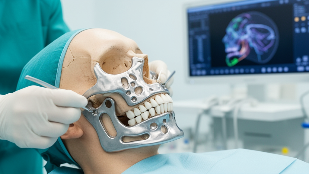
In a prospective single-arm clinical study investigating computer-assisted maxillofacial reconstruction, researchers found that combining virtual surgical planning with milled patient-specific titanium plates and autogenous split rib bundle bone grafts restored skeletal contour and facial symmetry in adults undergoing segmental mandibulectomy for benign jaw lesions. Segmental mandibular defects resulting from ablative surgery historically present profound reconstructive challenges, causing loss of facial symmetry, impaired mastication, speech disturbances, altered swallowing mechanics, and disruption of occlusal alignment. The trial evaluated whether preoperatively transferring digital three-dimensional surgical simulations to custom-milled fixation hardware provides a reproducible reconstructive alternative for patients presenting with benign pathologies who do not require vascularized free-flap tissue transfers. Across the cohort, the digital planning workflow facilitated graft adaptation and mandibular continuity without microvascular transfers.
The trial enrolled 30 adult patients presenting with benign mandibular lesions requiring immediate continuity restoration, with an average participant age of 37.23 ± 10.15 years. Among the participants, 17 individuals (56.7 percent) were male and 13 (43.3 percent) were female. Histopathological examination confirmed that ameloblastoma represented the most frequent surgical indication, diagnosed in 12 patients (40.0 percent), followed by odontogenic keratocyst in eight patients (26.7 percent), odontogenic myxoma in four patients (13.3 percent), vascular malformations in three patients (10.0 percent), and ossifying fibroma in three patients (10.0 percent). Surgical resections were categorized according to their anatomical extent: 13 patients (43.3 percent) had defects confined strictly to the mandibular body, nine patients (30.0 percent) had defects involving both the body and mandibular angle, and eight patients (26.7 percent) presented with defects spanning the body, angle, and ascending ramus. Strict eligibility criteria ensured that all candidates possessed adequate soft-tissue coverage and lacked prior or planned radiotherapy, active local infections, or recurrent lesions demanding wider composite soft-tissue resection.
The computer-assisted surgical workflow commenced with thin-slice computed tomography scans obtained from each patient, converted into three-dimensional digital mandibular models using specialized medical segmentation software. Utilizing Mimics and 3-matic software platforms from Materialise, the surgical team virtually performed intended resections, established osteotomy boundaries, and planned targeted postoperative mandibular alignment. To restore ideal anatomical contour, a virtual mirroring tool was applied against the unaffected contralateral mandibular anatomy to generate a customized plate model reproducing native bone curvature. The resulting digital hardware models were manufactured from medical-grade titanium (Grade 4) using milling techniques, producing custom plates with a uniform thickness of 1.5 millimeters. Notably, the clinical protocol relied on these patient-specific plates and anatomical alignment for surgical execution rather than using separate osteotomy, positioning, drilling, or costal harvest guides. This plate-assisted design aimed to establish correct segment positioning and eliminate the necessity for demanding intraoperative manual plate bending during surgery.
Surgeries were executed under general anesthesia through tailored approaches based on lesion location, combining intraoral lower sulcus incisions with extended extraoral submandibular access when required. When benign tumors extended to the mandibular angle, surgeons performed auxiliary coronoidectomies to minimize displacement of the proximal segment caused by temporalis muscle traction forces. For lesions involving or approximating the condyle, joint disarticulation was carried out while intentionally preserving the natural articular disc whenever feasible. The outer cortex of recipient stumps was decorticated using a rose-head bur to expose vascularized cancellous bone beds. Simultaneously, autologous rib grafts were harvested through a curved submammary incision, typically from two alternating ribs, preserving a costochondral component whenever condylar replacement was required, followed by layered donor wound closure upon confirming intact pleural integrity.
Harvested costal segments were split longitudinally along their axes using a fine osteotome to expose medullary marrow surfaces while preserving cancellous bone architecture. Fixation commenced by securing the milled 1.5-millimeter patient-specific titanium plate to residual mandibular segments using at least three screws on each side of the defect. One split rib segment was anchored proximally and a second distally within the prepared recipient beds. The remaining split rib pieces were telescoped to bridge the segmental gap, secured together as a bundle using screws with or without supplementary transosseous circumferential wires, and fixed directly to the titanium plate. Additional rib segments were added as necessary to restore vertical height, mesiodistal span, and buccolingual width while maintaining interarch clearance for future dental rehabilitation. The surgical team recorded a mean graft shaping and fixation time of 59.13 ± 12.25 minutes across the cohort, defined as the interval from rib harvest completion through final plate and graft fixation.
Radiographic evaluation demonstrated substantial improvements in skeletal symmetry when comparing preoperative baseline assessments against postoperative three-dimensional computed tomography measurements. The researchers quantified facial balance using a coordinate-based asymmetry index across five predefined skeletal landmarks: the condylion, lateral mandibular border, gonion, mental foramen, and midline menton. Across the cohort, the overall mean mandibular asymmetry index dropped from a preoperative baseline of 41.91 ± 5.32 millimeters to 6.48 ± 2.46 millimeters postoperatively, representing a statistically significant mean improvement of 35.43 millimeters (95% confidence interval, 33.63 to 37.23; p < 0.001). Individually, the gonion landmark exhibited the largest correction, declining from 49.33 ± 14.32 millimeters preoperatively to 8.38 ± 5.21 millimeters postoperatively (mean difference, 40.94; p < 0.001). Similarly, asymmetry at the condylion decreased from 37.99 ± 8.37 to 4.60 ± 4.12 millimeters, the lateral point improved from 38.90 ± 9.48 to 4.51 ± 4.36 millimeters, and the mental foramen improved from 41.41 ± 18.05 to 8.42 ± 5.53 millimeters, with all paired comparisons retaining significance following Holm-Bonferroni correction.
Postoperative anatomical alignment closely matched preoperative computer-generated surgical plans across the investigated anatomical landmarks. Radiographic discrepancy measurements between planned virtual positions and realized postoperative computed tomography coordinates demonstrated low spatial error, with mean deviations ranging from -0.003 ± 0.255 millimeters at the reconstructed mental foramen to 0.229 ± 0.383 millimeters at the unaffected contralateral gonion. Midline menton deviation exhibited a mean spatial difference of only 0.053 ± 0.266 millimeters relative to the virtual template. These imaging analyses were performed by a single examiner on anonymized, randomized computed tomography datasets, showing excellent intra-examiner reliability with intraclass correlation coefficients equal to or exceeding 0.99. However, the study authors explicitly noted that because titanium hardware and rib grafts remained visible on postoperative scans, complete examiner blinding was impossible. Furthermore, these evaluations relied on discrete anatomical landmarks rather than full surface-based three-dimensional registration, root-mean-square calculations, or surface distance mapping.
Serial volumetric assessment revealed progressive remodeling of nonvascularized split rib bundle grafts over the 12-month surveillance period. Semi-automated three-dimensional computed tomography segmentation demonstrated that mean graft volume across all 30 patients decreased from an immediate postoperative baseline of 15.61 ± 2.22 cubic centimeters to 14.60 ± 2.10 cubic centimeters at six months, reaching 14.11 ± 2.13 cubic centimeters at 12 months. Repeated-measures analysis of variance confirmed a statistically significant reduction in bone graft volume over time (Greenhouse-Geisser corrected F = 100.304, p < 0.001). Graft volume differed significantly among anatomical groups at every checkpoint: patients requiring body, angle, and ramus reconstruction started with an initial mean volume of 18.50 ± 0.69 cubic centimeters that settled to 16.86 ± 0.55 cubic centimeters at one year, while those with isolated body defects shifted from 13.53 ± 0.96 to 12.18 ± 1.09 cubic centimeters.
Clinical and functional evaluations indicated favorable short-term recovery alongside low postoperative morbidity. Stable dental occlusion was achieved and maintained in 28 patients (93.3 percent), while two patients (6.7 percent) presented with minor occlusal discrepancies that did not require secondary surgical correction; no frank malocclusion occurred. Overall graft survival reached 96.7 percent at 12 months, with 29 of 30 patients demonstrating solid osseous integration, continuous immobilization, and complete absence of graft exposure. Postoperative pain scores declined significantly across follow-up evaluations (Friedman test χ² = 88.946, p < 0.001), showing marked reductions between 24 hours, one week, and two weeks post-surgery. No instances of recipient-site or donor-site infection, wound dehiscence, occlusal relapse, or pneumothorax were observed. Only one patient (3.3 percent) experienced a late complication involving plate loosening and hardware removal that required secondary surgical intervention, which the authors hypothesized may have been related to suboptimal oral hygiene maintenance and local loading factors.
Subjective recovery was assessed through a five-point Likert scale, wherein 23 patients (76.7 percent) reported being very satisfied, four patients (13.3 percent) were satisfied, and three patients (10.0 percent) felt neutral, with no participants reporting dissatisfaction. The authors emphasized, however, that these findings reflect feasibility data from a single-arm trial conducted at a single specialized tertiary center without a comparative control cohort. Because the study did not include randomized or concurrent controls treated with standard non-custom plates, conventional freehand rib grafts, or microvascular free fibula flaps, the results cannot establish comparative superiority, equivalence, or non-inferiority against existing surgical techniques. Moreover, the 12-month follow-up remains insufficient to evaluate long-term graft remodeling, potential fatigue failure of titanium hardware, or definitive dental implant rehabilitation. The researchers concluded that while virtual surgical planning paired with patient-specific plates and split rib bundle grafting offers a viable reconstructive strategy for selected benign mandibular defects with healthy soft tissues, comparative trials with multi-observer accuracy analyses are needed to validate long-term clinical utility.
Subject of Research: Clinical and radiographic outcomes of virtual surgical planning and patient-specific plates in mandibular rib graft reconstruction.
Article Title: Short-term outcomes of virtual surgical planning–assisted mandibular reconstruction using a patient-specific reconstruction plate and split rib bundle bone graft: a prospective single-arm study
Article References: Abdeldayem, M., Hassan, S., Mohamed, M., & Shoukry, N. (2026). Short-term outcomes of virtual surgical planning–assisted mandibular reconstruction using a patient-specific reconstruction plate and split rib bundle bone graft: a prospective single-arm study. Maxillofacial Plastic and Reconstructive Surgery, 48(1), Article 21. https://doi.org/10.1186/s40902-026-00520-0
Image Credits: AI Generated
DOI: 10.1186/s40902-026-00520-0
Keywords: mandibular reconstruction, virtual surgical planning, patient-specific reconstruction plate, split rib bundle graft, segmental mandibulectomy, benign mandibular lesions, mandibular symmetry, asymmetry index, graft survival, computed tomography, cad cam technology, titanium plate, ameloblastoma, reconstructive surgery, maxillofacial surgery, graft volume, alexandria university, clinical feasibility
News Source: Ophelia Keating. (November 18, 2026). Virtual Planning and Patient-Specific Plates Achieve Mandibular Reconstruction in Feasibility Study. Scienmag.