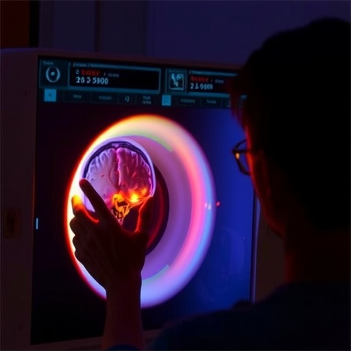
Researchers at the University of Virginia School of Medicine have embarked on a groundbreaking study backed by a $2.3 million grant from the U.S. Department of Defense, aiming to investigate the detection of subtle brain injuries in military personnel using advanced magnetic resonance imaging (MRI) technology. This pioneering research targets the long-standing challenge of visualizing microscopic brain damage in soldiers exposed to blast waves—a form of trauma traditionally invisible to conventional imaging techniques.
The focus of this initiative lies in identifying distinct brain scarring caused by astrocytes, specialized glial cells responsible for maintaining neural homeostasis and responding to injury. Prior neuropathological studies have detected astrocyte-driven scarring in soldiers’ brains post-mortem, revealing changes linked to repeated blast exposures during military activities. However, such insights were limited to histological examination after death, leaving a critical gap in diagnosing living patients afflicted with similar injuries.
To bridge this gap, the new MRI scanner installed at UVA’s Fontaine Research Park leverages cutting-edge imaging sequences and hardware to enhance sensitivity to microstructural and biochemical changes in brain tissue. These novel imaging protocols are designed to capture subtle astrocytic scarring and related pathological alterations, which elude standard clinical MRI scans. The scanner’s potential to reveal these minuscule but consequential brain injuries promises to revolutionize diagnostic practices in military medicine.
The implications of successfully visualizing blast-induced brain scarring are profound. First, it offers a means to diagnose service members whose routine brain scans currently appear normal, yet who suffer debilitating cognitive and neurological symptoms. This advanced detection capability paves the way toward personalized treatment strategies by mapping the extent and location of brain lesions, tailoring rehabilitative or pharmacological interventions accordingly.
Furthermore, the enhanced MRI technology stands to accelerate research into neuroprotective therapies targeting blast-related neuropathology. By providing objective imaging biomarkers, clinicians can more effectively evaluate therapeutic efficacy in clinical trials, expediting the development of treatments to mitigate chronic neurological deficits observed in blast-exposed individuals. Such progress could dramatically improve the quality of life for veterans and active-duty personnel alike.
Understanding the threshold and cumulative effects of blast exposure represents another crucial objective of this study. Military personnel are frequently subjected to low-level blasts during training and combat, with mounting evidence linking these exposures to progressive brain injury. The advanced MRI technology could yield quantitative measures correlating blast intensity and frequency with the degree of brain injury, informing safer operational protocols and protective measures for service members.
The research project, slated for a three-year duration, will enroll 60 service members exhibiting a spectrum of blast exposure histories. Participants will undergo comprehensive neuroimaging alongside rigorous neuropsychological assessments aimed at evaluating cognitive, emotional, and behavioral functions. Integrating imaging and clinical data, the researchers hope to delineate the relationship between blast-induced structural brain changes and clinical symptomatology.
This multidisciplinary endeavor is led by Dr. James R. Stone, MD, PhD, a radiologist with expertise in neuroimaging and blast injury pathology. Dr. Stone collaborates closely with colleagues from the Naval Medical Research Command and other military-affiliated academic health centers. Together, their concerted efforts align with the objectives of UVA’s newly inaugurated Paul and Diane Manning Institute of Biotechnology, which prioritizes translational research with tangible clinical impact.
Blast-induced neurotrauma represents an insidious threat to service members’ health, with repeated low-level exposures contributing to cumulative brain damage that evades immediate detection. Unlike overt traumatic brain injuries resulting from blunt force trauma, blast injuries often manifest as diffuse axonal injury and astrocytic scarring—processes that produce subtle but long-lasting cognitive impairments, emotional disturbances, and neuropsychiatric disorders.
Conventional imaging modalities, including CT and standard MRI scans, lack the resolution and specificity required to detect these microscopic alterations. The technological advancements embodied in the innovative MRI system under study incorporate diffusion tensor imaging, susceptibility-weighted imaging, and other sophisticated sequences designed to elucidate microstructural integrity, iron deposition, and metabolic perturbations within affected brain regions.
Beyond diagnostic enhancement, the project aspires to deepen fundamental scientific understanding of blast neuropathology. The correlation of imaging findings with neuropsychological profiles will provide unprecedented insights into the mechanisms by which astrocyte-driven scarring impacts neural circuitry, synaptic function, and cognitive processing. This knowledge is critical for crafting targeted interventions that not only manage symptoms but also halt or reverse underlying neuropathological progression.
The potential societal benefits of this research extend well beyond the military community. Civilian populations exposed to blast injuries in industrial accidents, terrorist attacks, or other contexts could similarly benefit from refined imaging diagnostics and tailored therapeutic approaches informed by this study’s findings. Thus, the project stands at the intersection of military medicine, neuroscience, and public health innovation.
Ultimately, the success of this advanced imaging approach in detecting previously hidden brain injuries could redefine clinical paradigms governing the management of blast-related neurotrauma. Early diagnosis, precise monitoring, and individualized treatment strategies enabled by this technology will help ensure service members receive timely care, mitigate long-term disability, and improve reintegration outcomes after exposure to hazardous environments.
As the study progresses, continuous dissemination of discoveries, methodologies, and clinical guidelines will be essential to foster widespread adoption of these innovations across defense and civilian healthcare systems. This endeavor exemplifies how scientific rigor, technological advancement, and military medicine collaboration converge to address vexing healthcare challenges with profound human and societal implications.
Subject of Research: Advanced MRI detection of blast-induced brain injuries in military personnel
Article Title: Cutting-Edge MRI Technology Aims to Reveal Hidden Brain Scarring in Blast-Exposed Soldiers
News Publication Date: Not specified
Web References: https://manninginstitute.virginia.edu/ ; https://makingofmedicine.virginia.edu/
Image Credits: UVA Health
Keywords: Brain damage, Traumatic injury, Head concussions, Neuroprotection
Tags: advanced MRI technologyastrocyte-driven brain scarringblast wave trauma effectsbrain injury detection in soldiersDepartment of Defense funding researchglial cells and neural homeostasisinnovative neuroimaging techniquesliving patient brain injury diagnosismicroscopic brain damage imagingmilitary personnel brain healthsubtle brain injury identificationUVA MRI studies


