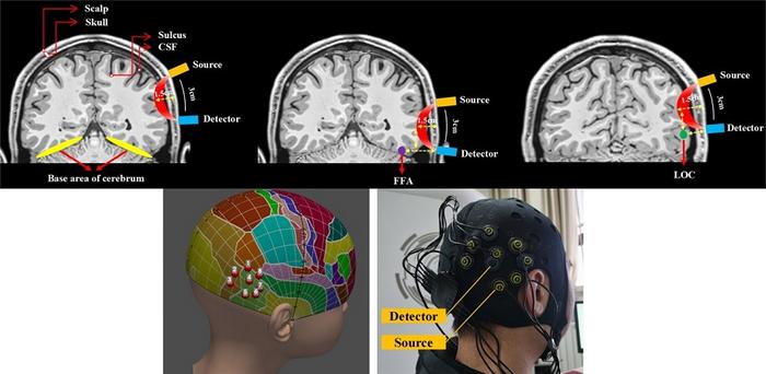
The brain is not only the most complex organ of the human body, but also one of the most difficult to study. To understand the roles of different regions of the human brain and how they interact, it is crucial to measure neuronal activity with awake subjects while they perform controlled tasks. However, the most accurate measurement devices are invasive, which greatly limits their use on healthy humans in real-life settings.

Credit: W. Chai, P. Zhang, et al., doi 10.1117/1.NPh.11.1.015002.
The brain is not only the most complex organ of the human body, but also one of the most difficult to study. To understand the roles of different regions of the human brain and how they interact, it is crucial to measure neuronal activity with awake subjects while they perform controlled tasks. However, the most accurate measurement devices are invasive, which greatly limits their use on healthy humans in real-life settings.
To overcome this major obstacle, scientists have come up with ingenious techniques to measure brain activity in safe and noninvasive ways. One prominent example is functional magnetic resonance imaging (fMRI), which uses strong magnetic fields and radio waves to map changes in blood flow in the brain. A major drawback of fMRI is the size and cost of the necessary equipment, which restricts its more widespread adoption in laboratory and clinical settings. Fortunately, a different technique called functional near-infrared spectroscopy (fNIRS) has been gaining traction. This method involves placing a light source and detector on the scalp to measure localized changes in hemoglobin concentration, which correlate to brain activity. Despite its advantages, which include simplicity and portability, the true potential of fNIRS remains unexplored in many regions of the brain.
Previous studies have used fNIRS to detect brain activity in the ventral visual pathway, but none have evaluated its feasibility and ecological validity, or whether the signal detected is desirable. Against this backdrop, a research team including Professor Minghao Dong from Xidian University, China, together with Professor Chaozhe Zhu from Beijing Normal University, set out to test the capabilities of fNIRS for measuring brain activity in the lateral occipital complex (LOC) and the fusiform face area (FFA), two key regions in what is known as the ventral visual pathway. Their study is published in the Gold Open Access journal Neurophotonics.
To understand the experiments, it helps to know the functions of the LOC and FFA. The LOC plays a crucial role in object recognition; its neurons are involved in processing information about the shapes and forms of objects. On the other hand, the FFA is specialized in the processing and recognition of faces. Compared to the FFA, the LOC is much closer to the scalp. Thus, the team hypothesized that fNIRS measurements were more likely to be successful in this region than on the LOC.
To test this hypothesis, the researchers recruited 63 adult subjects, of which 35 were included in the current study, while the other 28 were included in a follow-up study, the results of which matched those in the current study but were not mentioned in the publication. The team conducted several object- and face-recognition tasks while performing fNIRS measurements using a portable instrument. The idea was to check if the corresponding brain region would exhibit activity in response to images of objects or faces that the subject had seen previously during the experiment. Worth noting, the team employed a tool called the “transcranial brain atlas,” which they had developed in a previous study, to determine the optimal placement of the instrument’s sensors for each individual subject. Moreover, the current study provides valuable insights by demonstrating that placing the target channel corresponding to the target coordinates is sufficient for measuring LOC activity, eliminating the need for additional supplementary channels around the target coordinates.
The results matched the researchers’ expectations, as Dong remarks: “According to our findings, the LOC target channel selectively activates in reaction to objects, whereas the FFA target channel does not.” The most likely explanation is that the depth at which the FFA is located exceeds the detection threshold of fNIRS. “The LOC region appears to be a suitable target for fNIRS-based detection, despite the fact that fNIRS detection has limitations in collecting FFA activity,” adds Dong.
Overall, the research team’s efforts represent a step towards better techniques with which to study the brain. “Our results help understand the feasibility of fNIRS for practical applications. To the best of our knowledge, this work is the first to examine the viability of the technique in monitoring cortical activity within the ventral visual pathway,” concludes Dong.
Further advances in fNIRS technology could lead to practical, low-cost diagnostics for certain brain disorders, as well as potential avenues for neuroenhancement devices. Such equipment would enable us to augment specific cognitive functions or help treat neurological conditions.
Only time will tell what fNIRS may hold for the future of neuroscience!
For details, see the original article by Chai, Zhang, et al., “Feasibility study of functional near-infrared spectroscopy in the ventral visual pathway for real-life applications,” Neurophotonics 11(1) 015002 (2024), doi 10.1117/1.NPh.11.1.015002.
Journal
Neurophotonics
DOI
10.1117/1.NPh.11.1.015002
Article Title
Feasibility study of functional near-infrared spectroscopy in the ventral visual pathway for real-life applications
Article Publication Date
8-Jan-2024


