In a groundbreaking study that could redefine our understanding of mammalian reproduction, researchers have unveiled the intricate molecular structure of the cytoplasmic lattice (CPL) within oocytes. The CPL, a fibrous network present in the cytoplasm of mammalian oocytes, plays a pivotal role in oocyte maturation and the earliest stages of embryonic development. Despite its well-known presence since the 1960s, the molecular intricacies and assembly mechanics of the CPL had remained largely elusive—until now.
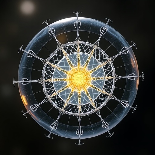
Utilizing cutting-edge cryo-electron microscopy (cryo-EM), the research team has provided an unprecedented glimpse into the CPL’s molecular architecture, specifically in mouse oocytes. This advanced imaging enabled the identification of 14 distinct protein subunits that collectively construct the lattice’s complex framework. These findings illuminate how the CPL’s repeating structural units assemble into a highly organized filamentous network critical for developmental processes.
At the heart of the CPL’s architecture are two fundamental motifs—dubbed the “U-shaped basket” (UB) and the “adapter ring” (AR). These repeating units interlock with remarkable precision to form the CPL’s filamentous backbone. The adapter ring exhibits a two-fold symmetry, integrating key proteins including NLRP4f, SCMC components, and ZBED3. The AR’s circular assembly relies on two interaction clusters, which stabilize its structure and facilitate connections between adjacent CPL units.
Anchoring the U-shaped basket is the enzyme PADI6, a particularly notable protein that has been previously implicated in oocyte quality and embryonic competence. PADI6 forms a didecameric complex made up of ten homodimers arranged as two back-to-back pentamers. These pentamers constitute the lateral sides of the UB structure, providing essential scaffolding that supports the entire CPL framework.
The base and vertical sides of the UB are further elaborated by multisubunit complexes exhibiting central symmetry. On the basal side, a complex comprising UBE2D3, UHRF1, and NLRP14 proteins crafts a stable foundation, while the vertical sides are formed by a combination of tubulin isoforms TUBB2B and TUBB2A, along with FBXW24 and SKP1. These intricate assemblies interface directly with the PADI6 pentamers, collectively consolidating the UB’s stability and shape.
Integral to maintaining the continuous filamentous lattice are connections mediated by SCMC dimers embedded within each adapter ring. These dimers bridge the vertical facets of two neighboring UBs, fostering a dense protein-protein interaction network. This repetitive linkage acts as the molecular glue, ensuring CPL units are cohesively joined while preserving the lattice’s periodic structure.
This discovery transcends prior knowledge, revealing how large, periodic filamentous structures like the CPL are molecularly orchestrated within mammalian oocytes. The elaborate protein network not only provides mechanical support but likely coordinates key biochemical events required during the oocyte-to-embryo transition.
The implications of understanding CPL assembly extend beyond foundational cell biology. Given that CPL integrity correlates with early embryonic developmental competence, these insights could have profound impacts on reproductive medicine. Defects in CPL formation or function might underlie certain female infertility disorders, opening avenues for novel diagnostic and therapeutic strategies.
Moreover, the study highlights PADI6 as a central architect of CPL formation, reaffirming its significance as a critical maternal effect protein. Targeting PADI6 interactions or modulating its assembly pathway could become a strategic focus for enhancing oocyte quality in assisted reproductive technologies (ART).
This research also exemplifies how cryo-EM is revolutionizing the visualization of complex cellular structures in near-native states, enabling discoveries that were previously inaccessible. The resolution achieved in delineating the CPL’s protein constituents sets a new benchmark for studies of mesoscale biomolecular assemblies.
Future investigations inspired by this work will likely explore how dynamic changes in CPL architecture regulate the timing of oocyte maturation and embryo activation. Understanding these processes at atomic resolution paves the way for deciphering the molecular crosstalk between CPL components and other cellular machineries.
In sum, this comprehensive elucidation of the CPL’s structural blueprint unlocks a fundamental aspect of mammalian developmental biology. It serves as a molecular Rosetta stone, translating the enigmatic cytoplasmic lattice into an organized, functional filament network intricately woven by multiple protein complexes.
As the first detailed molecular map of the CPL emerges, it sets the stage for exploring how perturbations in this lattice influence cellular health and reproductive outcomes. The knowledge gained not only enriches our basic scientific understanding but also holds promise for addressing critical challenges in fertility medicine and embryology.
Liu et al.’s landmark study thus represents a monumental leap forward, bridging decades of cytological observations with state-of-the-art molecular visualization. Their findings provide a crucial foundation for future interventions aimed at safeguarding early mammalian development and female reproductive wellness.
Subject of Research: Mammalian oocyte cytoplasmic lattice (CPL) assembly and molecular architecture
Article Title: Molecular basis of oocyte cytoplasmic lattice assembly
Article References:
Liu, S., Liu, Y., Xue, J. et al. Molecular basis of oocyte cytoplasmic lattice assembly. Nature (2026). https://doi.org/10.1038/s41586-026-10360-7
Image Credits: AI Generated
Tags: adapter ring structure in oocyte CPLCPL role in embryonic developmentcryo-electron microscopy in oocyte researchfilamentous network in oocyte cytoplasmmammalian oocyte maturation mechanismsmolecular assembly of cytoplasmic latticeNLRP4f protein in oocyte latticeoocyte cytoplasmic lattice structureprotein subunits in oocyte CPLSCMC complex in oocyte cytoplasmU-shaped basket motif in CPLZBED3 protein function in CPL
