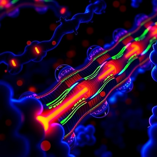
In a groundbreaking advancement in ion channel research, scientists have unveiled the molecular underpinnings of fast N-type inactivation in voltage-gated potassium (K_v) channels, a fundamental process that regulates electrical signaling in excitable cells. Using state-of-the-art cryo-electron microscopy (cryo-EM), the team has resolved near-atomic structures of the Shaker K_v channel in unprecedented detail, shedding light on how specific N-terminal peptides mediate rapid channel inactivation. This revelation offers critical insights into the dynamic gating mechanisms that control neuronal firing and muscle contraction, with profound implications for understanding electrical disorders and developing targeted therapeutics.
The Shaker K_v channel, originally identified in Drosophila melanogaster, has long served as a canonical model for studying voltage-gated potassium channels. These channels are pivotal in shaping action potentials and repolarizing the membrane following neuronal excitation. Of particular interest is the fast N-type inactivation mechanism, whereby the channel’s N-terminal “ball peptide” swiftly occludes the pore, halting potassium flow within milliseconds. Despite decades of electrophysiological characterization, the structural basis for this rapid inactivation remained elusive due to the intrinsic flexibility and transient nature of the involved protein domains.
.adsslot_okUpG9v4mr{width:728px !important;height:90px !important;}
@media(max-width:1199px){ .adsslot_okUpG9v4mr{width:468px !important;height:60px !important;}
}
@media(max-width:767px){ .adsslot_okUpG9v4mr{width:320px !important;height:50px !important;}
}
ADVERTISEMENT
Crucially, subsequent reconstitution into near-native lipid nanodiscs composed of defined phospholipid mixtures mimicked the physiological membrane environment, enabling high-resolution structural studies. A meticulously calibrated molar ratio of the Shaker tetramer to membrane scaffold proteins and lipids was employed, ensuring optimal incorporation and functional preservation. This lipid nanodisc platform was pivotal for visualizing biologically relevant conformations that detergents alone cannot sustain, highlighting the interplay between lipids and channel gating.
Cryo-EM grids were prepared under carefully controlled conditions, including the strategic use of fluorinated Fos-choline-8 detergent to enhance particle distribution and orientation. Data acquisition leveraged a Titan Krios microscope equipped with a Gatan K3 direct electron detector at super-resolution mode, yielding remarkable image quality. Processing pipelines combined powerful software tools such as RELION and cryoSPARC, employing sophisticated motion correction, contrast transfer function estimation, and particle classification strategies. Notably, symmetry expansion and focused classification with masks targeting the internal pore and surrounding chambers enabled distinguishing multiple functional states from over a million particles.
The resulting high-resolution maps revealed intricate details of the N-terminal peptide’s engagement within the channel pore. The “ball” region extended deep into the internal vestibule, adopting an L-shaped density pattern that pinpoints key residues responsible for rapid occlusion. Complementary density corresponding to the T1 domain, isolated through subtraction and dedicated refinement, attained sub-3 Å resolution, providing architectural insights into channel tetramerization and the spatial context of the N-terminal peptide.
Parallel mass spectrometry analyses affirmed the presence and post-translational modifications of the N-terminal regions, bolstering the structural interpretations. Electrophysiological validation employed Xenopus laevis oocytes injected with mRNA or liposome-reconstituted channels, probing voltage-dependent activation and inactivation kinetics. The team dissected the voltage sensitivity and charge movements underlying gating transitions by fitting Boltzmann functions to conductance-voltage relationships, further clarifying the biophysical consequences of the observed structural motifs.
Intriguingly, mutant constructs such as E12K/D13K variants displayed altered inactivation behaviors, correlating with disruptions in the peptide’s pore-binding mode captured in cryo-EM. These observations offer mechanistic explanations for mutations affecting channelopathies in humans and suggest avenues for modulating channel inactivation through targeted interventions. The meticulous combination of structural, biochemical, and electrophysiological data underscores the complexity and finely tuned nature of fast inactivation processes.
This study exemplifies how convergent methodologies can unravel dynamic, small-domain interactions previously intractable to structure determination. The elucidation of the fast N-type inactivation gate architecture in a voltage-gated K+ channel propels our understanding of ion channel regulation and sets the stage for exploring similar mechanisms in other channel families. Moreover, the insights gleaned provide a molecular blueprint that can inform drug design efforts targeting hyperexcitability disorders such as epilepsy, neuropathic pain, and cardiac arrhythmias.
Future directions opened by this work include time-resolved cryo-EM studies to capture the kinetics of the inactivation process, as well as investigations into how lipid composition and membrane tension influence channel gating. The remarkable resolution achieved also invites exploration of subtler conformational states and allosteric modulatory sites. Beyond fundamental biology, this research heralds a new era where the integration of structural and functional techniques enables rational engineering of ion channels with bespoke properties for therapeutic and synthetic biology applications.
In summary, by harnessing advanced structural biology techniques and functional assays, researchers have cracked the enigma of rapid N-type inactivation in the Shaker K_v channel. The detailed visualization of the N-terminal peptide’s pore-blocking conformation not only vindicates longstanding electrophysiological models but also reveals new molecular intricacies. This landmark achievement enhances our comprehension of neuronal excitability regulation and offers a potent platform for drug discovery targeting ion channel dysfunction.
Subject of Research: Structural and functional characterization of fast N-type inactivation mechanism in voltage-gated Shaker K_v channels.
Article Title: Structural basis of fast N-type inactivation in K_v channels
Article References:
Tan, XF., Fernández-Mariño, A.I., Li, Y. et al. Structural basis of fast N-type inactivation in K_v channels. Nature (2025). https://doi.org/10.1038/s41586-025-09339-7
Tags: cryo-electron microscopy applicationsDrosophila melanogaster ion channelsdynamic gating mechanisms in K_v channelselectrical signaling in excitable cellsfast N-type inactivationmolecular underpinnings of ion channelsmuscle contraction regulationneuronal firing mechanismsprotein engineering in ion channel researchShaker Kv channel structuretargeted therapeutics for electrical disordersvoltage-gated potassium channels


