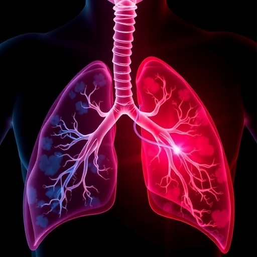
In the realm of neonatal medicine, bronchopulmonary dysplasia (BPD) remains a formidable challenge, particularly affecting premature infants with devastating effects on lung development and function. This chronic respiratory condition is more than just a consequence of early birth; it entails a complex interplay of cellular and molecular disruptions that ultimately sculpt the long-term pulmonary landscape for these vulnerable newborns. Recent advances in bioinformatics have opened new avenues in understanding the underpinnings of BPD, specifically highlighting the pivotal role of mitophagy, a selective form of autophagy responsible for mitochondrial quality control. A groundbreaking study published in Pediatric Research by Li, Wang, Wang, and colleagues delves deeply into this mitochondrial dynamic, unravelling critical molecular pathways implicated in the pathogenesis of BPD.
At the core of BPD’s pathology lies the disturbance of normal lung development, characterized by impaired alveolarization and vascular growth. These developmental aberrations are compounded by persistent inflammation and oxidative stress, further exacerbated by the immature antioxidant defenses of preterm infants. Within this context, mitochondrial dysfunction emerges as a central contributor to cellular damage and inflammation in the lung tissue. Mitochondria, the powerhouses of the cell, also play integral roles in signaling and apoptosis; their selective degradation through mitophagy ensures cellular homeostasis by removing damaged or dysfunctional mitochondria. This process is crucial during the heightened oxidative stress conditions observed in premature lungs subjected to mechanical ventilation or oxygen therapy.
Leveraging the power of bioinformatics, Li and colleagues conducted comprehensive transcriptomic analyses to identify key regulators of mitophagy in lung tissues affected by BPD. By integrating high-throughput sequencing data and advanced computational algorithms, they were able to map intricate gene expression profiles that correlate with mitophagy activity. This approach uncovered a previously unappreciated landscape of mitophagic dysregulation, providing clues about specific molecules and pathways that may either exacerbate or mitigate lung injury in BPD. Importantly, these findings underscore the potential for mitophagy modulation as a therapeutic strategy in neonatal care.
.adsslot_I9Wztq0lnj{ width:728px !important; height:90px !important; }
@media (max-width:1199px) { .adsslot_I9Wztq0lnj{ width:468px !important; height:60px !important; } }
@media (max-width:767px) { .adsslot_I9Wztq0lnj{ width:320px !important; height:50px !important; } }
ADVERTISEMENT
One of the striking revelations from the study was the identification of several mitophagy-related genes whose expression patterns were significantly altered in BPD. Genes encoding proteins involved in the recognition and removal of damaged mitochondria, such as PINK1 and Parkin, showed dysregulated expression. The perturbation of these genes suggests a compromised mitochondrial quality control mechanism, which may lead to accumulation of defective mitochondria, escalating oxidative stress and triggering inflammatory cascades that damage the delicate lung parenchyma. Such molecular insights provide a more granular understanding of how cellular energy metabolism intertwines with inflammatory responses in BPD.
Furthermore, Li et al.’s work highlights the interconnectedness between mitophagy and other cellular processes implicated in lung injury. For instance, the interplay between mitophagy and endoplasmic reticulum (ER) stress was particularly prominent. ER stress has been known to induce inflammatory signaling and apoptosis, and dysfunctional mitophagy can amplify ER stress, creating a vicious cycle that impairs lung cell survival and regeneration. By revealing these complex interdependencies, the study contributes to a holistic view of the cellular milieu in BPD, offering new targets for clinical intervention.
The methodological rigor of this study is grounded in meticulous data curation and sophisticated analytics. The team employed integrative bioinformatics tools to analyze gene ontology and pathway enrichment, revealing that altered mitophagy genes were often involved in pathways related to immune responses, oxidative stress, and cell death. This multifaceted impact underscores mitophagy’s role as a molecular hub in BPD pathogenesis, where its dysfunction leads to widespread effects across cellular systems that govern lung development and immune homeostasis.
Perhaps one of the most promising aspects of this research is its translational potential. By profiling mitophagy mechanisms at a molecular level, the study paves the way for developing biomarker-driven diagnostics that can identify infants at higher risk for severe BPD. Moreover, it opens up possibilities for therapeutic interventions aimed at restoring mitophagy balance. Pharmacological agents capable of enhancing mitophagy could, theoretically, mitigate mitochondrial damage and dampen the inflammatory milieu in the immature lung, potentially improving clinical outcomes for preterm infants facing this debilitating condition.
The implications of this study extend beyond the lungs, as the systemic effects of mitophagy dysfunction may influence other organs impacted by prematurity and oxygen toxicity. The intricate crosstalk between mitochondrial dynamics and immune modulation suggests that mitophagy-targeted therapies could confer benefits by addressing multi-organ vulnerabilities in preterm infants. Such approaches would represent a paradigm shift in neonatal intensive care, moving from symptomatic treatment towards mechanistically informed strategies.
Interestingly, this research also raises intriguing questions about the temporal dynamics of mitophagy in BPD progression. Understanding whether mitophagy impairment is an early event that predisposes to lung injury or a secondary consequence of established pathology is critical for timing therapeutic interventions. Future studies might focus on longitudinal monitoring of mitophagy markers in newborns, complemented by animal models that recapitulate the human BPD phenotype, to elucidate causality and therapeutic windows.
In addition, the study’s reliance on bioinformatic analyses exemplifies the power of big data in neonatal research. As large-scale omics datasets become increasingly available, integrating multi-dimensional data—genomic, proteomic, metabolomic—will be vital for constructing comprehensive molecular maps of diseases like BPD. Such interdisciplinary approaches promise to unravel complexities that are invisible to traditional experimental methods, accelerating discovery and innovation in pediatric medicine.
The work by Li and colleagues also highlights the critical role of collaborative research, combining clinical insights with computational biology expertise. This synergy is paramount for tackling multifactorial diseases where the interaction of genetic, environmental, and therapeutic factors create intricate pathological networks. By bridging these disciplines, the study sets a precedent for future investigations into neonatal diseases characterized by mitochondrial dysfunction and oxidative stress.
Understanding the molecular choreography of mitophagy in bronchopulmonary dysplasia could redefine the clinical management of this condition. Identification of safe and effective mitophagy modulators will require rigorous preclinical testing and carefully designed clinical trials. However, the foundational knowledge provided by this study is an essential step toward personalized medicine in neonatology, whereby interventions are tailored to molecular phenotypes rather than broad clinical symptoms.
In conclusion, this comprehensive bioinformatics investigation into mitophagy’s role in bronchopulmonary dysplasia represents a landmark in neonatal respiratory research. By illuminating the molecular dysfunctions centered on mitochondrial quality control, the study not only deepens fundamental understanding of BPD pathogenesis but also inspires new directions for clinical innovation. The convergence of computational biology and neonatology embodied in this work showcases the future of precision medicine strategies aimed at the earliest stages of human life.
Subject of Research: Bronchopulmonary dysplasia and the role of mitophagy in its molecular pathogenesis.
Article Title: Investigating mitophagy mechanisms in bronchopulmonary dysplasia through bioinformatics.
Article References:
Li, C., Wang, Y., Wang, X. et al. Investigating mitophagy mechanisms in bronchopulmonary dysplasia through bioinformatics. Pediatr Res (2025). https://doi.org/10.1038/s41390-025-04319-z
Image Credits: AI Generated
DOI: https://doi.org/10.1038/s41390-025-04319-z
Tags: alveolarization in lung developmentantioxidant defenses in preterm infantsbioinformatics in neonatal medicinebronchopulmonary dysplasia in neonatescellular and molecular pathways in BPDinflammation in bronchopulmonary dysplasiamitochondrial dysfunction in BPDmitophagy and lung developmentneonatal respiratory conditionsoxidative stress in premature infantspediatric respiratory health challengesselective autophagy mechanisms

