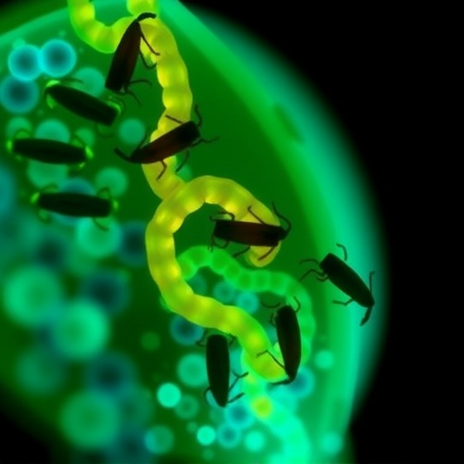
In a groundbreaking advancement poised to reshape the study of lipid biology in small organisms, a collaborative team of scientists from Japan and the Netherlands has unveiled a pioneering method for high-resolution, three-dimensional lipid imaging in the nematode Caenorhabditis elegans. This technique, combining the precision of matrix-assisted laser desorption/ionization mass spectrometry imaging (MALDI-MSI) with innovative microfluidics and traditional histological staining, offers an unprecedented view of fat distribution at the organ-specific level within this widely studied model organism.
For decades, lipid research in C. elegans has been limited by technological hurdles. While fat storage and metabolism are central to understanding aging, disease, and metabolic regulation, existing methods failed to simultaneously provide molecular identity and spatial context for lipids inside intact tissues. Typically, researchers relied either on lipid-specific stains that lack molecular specificity or on mass spectrometry techniques that lost anatomical information through sample degradation or preparation constraints. This new workflow meticulously overcomes these challenges by preserving the worm’s internal architecture while revealing intricate lipid localization.
.adsslot_AhU1itDRGo{ width:728px !important; height:90px !important; }
@media (max-width:1199px) { .adsslot_AhU1itDRGo{ width:468px !important; height:60px !important; } }
@media (max-width:767px) { .adsslot_AhU1itDRGo{ width:320px !important; height:50px !important; } }
ADVERTISEMENT
This multi-modal approach grants researchers the ability to spatially correlate molecular lipid identity with anatomical features in ways previously unattainable. Notably, the research group produced a series of ten consecutive cross-sections from individual nematodes, enabling digital reconstruction of 3D lipid maps across the entire ~1 mm-long organism. These maps reveal remarkable heterogeneity in lipid presence and concentration across different tissues, including the pharynx, intestine, and reproductive organs. For example, lipids connected to cholesterol metabolism predominantly localized to the pharynx and anterior intestinal regions, suggesting functional roles in nutrient uptake and processing.
The preservation of the nematode’s microanatomy during analysis proved critical. Unlike conventional lipid profiling methods that sacrifice either spatial context or molecular specificity, this framework maintains sub-organ resolution, which is essential for dissecting the complex interactions between lipid metabolism and cellular physiology. The data demonstrated high reproducibility, with biological variability between individual worms surpassing any technical noise, underscoring the method’s robustness and reliability.
Beyond its technical merits, this methodology represents a strategic leap for biomedical research using C. elegans as a proxy. Given the worm’s genetic and physiological parallels to higher organisms, insights into tissue-specific lipid dynamics can illuminate mechanisms underlying human metabolic disorders, neurodegeneration, and age-related diseases. The ability to monitor lipid alterations in response to genetic mutations, pharmacological interventions, or environmental stresses positions this technique as a formidable tool in translational research.
Dr. Masazumi Fujiwara, leading the study from Okayama University’s Nanochemistry Laboratory, emphasizes that this integrated approach opens new avenues for quantitative and qualitative lipid research in invertebrate models. His PhD student, Sara Mandic, who spearheaded the imaging efforts, remarks on the breakthrough potential of this technology: “For the first time, we can map distinct lipid molecules with precise spatial resolution inside an entire live organism without compromising its structural context.” The study exemplifies the synergy achievable by blending engineering, chemistry, and biology.
Experimentally, the team overcame several technical barriers to optimize sample preparation and imaging. The delicate process of embedding nematodes into stabilizing matrices while preventing lipid diffusion or degradation presented a significant challenge. The microfluidic chip’s design ensures consistent orientation and immobilization, crucial for acquiring serial sections that seamlessly align for 3D reconstruction. MALDI-MSI parameters were finely tuned to enhance sensitivity and spatial resolution, enabling detection of a broad range of lipid species, including phospholipids, glycolipids, and sterols.
The combination of MALDI-MSI with histological staining sets a new standard for correlating molecular profiles with classical anatomy. Oil Red O stain, long used for detecting neutral triglycerides and cholesteryl esters, anchors the molecular signals detected by MALDI to familiar histological landmarks. This dual visualization fosters a deeper understanding of lipid function in contexts such as lipid storage droplets versus membrane-associated lipids, enhancing interpretive clarity.
Importantly, the study’s reproducibility across individual nematodes ushers in robust comparative analyses, allowing researchers to explore how lipid distributions vary with developmental stages, genetic backgrounds, or environmental exposures. This will facilitate identification of lipid biomarkers indicative of physiological changes or disease phenotypes, accelerating discovery pipelines in aging and metabolic research.
Looking forward, the team plans to expand this technique’s utility by integrating quantitative lipidomics and applying it to disease models within C. elegans. The method’s compatibility with genetically modified strains holds promise for unraveling how specific genes regulate lipid metabolism and distribution in vivo. Moreover, coupling this workflow with emerging bioinformatics tools could enable high-throughput spatial lipidomics, yielding comprehensive atlases of lipid networks in health and disease.
The study’s implications transcend C. elegans research alone. The demonstrated ability to preserve anatomical fidelity while acquiring detailed molecular data represents a conceptual advance applicable to diverse small organisms and possibly mammalian tissues. Such integrative spatial omics techniques are poised to revolutionize biomedical sciences by providing holistic views of biomolecular organization that underpin physiology and pathology.
This unprecedented 3D lipid imaging modality not only enriches our fundamental knowledge of invertebrate biology but also equips scientists with critical insights into the tissue-specific roles of lipids in metabolic regulation. In a landscape where metabolic disorders and age-related diseases are rising global health concerns, breakthroughs of this nature offer hope for identifying novel therapeutic targets informed by molecular and spatial precision.
Altogether, the multidisciplinary effort led by Okayama University and Maastricht University embodies a formidable stride towards dissecting lipid biology with unparalleled clarity. As this technology is adopted and refined, it promises to catalyze discoveries that interlink molecular lipidomics with cellular anatomy, ultimately deepening our understanding of fat’s multifaceted roles in life and disease.
Subject of Research: Animals
Article Title: Method development for correlating lipid molecular information with anatomy in C. elegans
News Publication Date: 8-Jul-2025
Web References:
https://doi.org/10.1038/s41598-025-09577-9
Image Credits:
Sara Mandic from Okayama University, Japan
Keywords:
Life sciences, Lipids, Cell biology, Biochemistry, Histological analysis, Developmental biology, Molecular biology, Embryos, Mass spectrometry
Tags: collaborative research in lipid sciencefat distribution analysis in nematodeshigh-resolution lipid imaging techniqueinnovative methods in lipid biologylipid metabolism and aging studiesMALDI mass spectrometry imaging C. elegansmicrofluidics in lipid researchnematode model organism advancementsorgan-specific lipid localization techniquesovercoming technological hurdles in lipid studiesspatial context in lipid researchtissue architecture preservation in mass spectrometry

