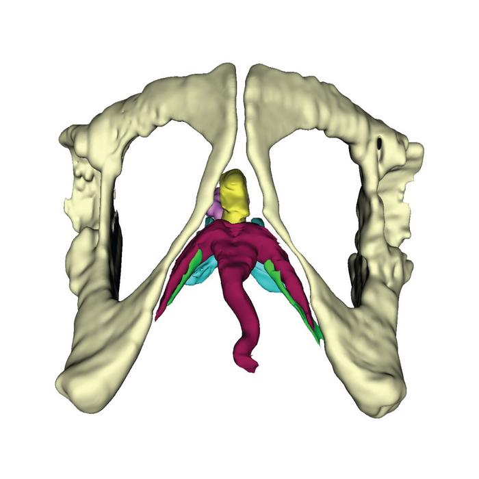
New research into the anatomy of the primate clitoris using sophisticated imaging technology reveals the wild variation in clitoral form between primate species.

Credit: Dr Varajão de Latorre
New research into the anatomy of the primate clitoris using sophisticated imaging technology reveals the wild variation in clitoral form between primate species.
Despite its popularity, the clitoris remains widely understudied scientifically and there is still much to learn about its anatomical form and function. This scarcity of research is even more pronounced for other animals, including our closest living relatives, the non-human primates, a group that includes apes, monkeys, lemurs and tarsiers.
“The subsurface 3D shape of the human clitoris was only fully described in the last 20 years, despite us having equivalent information on male genitalia for much longer,” says Dr Daniel Varajão de Latorre, a post-doctoral researcher from the Manchester Metropolitan University, UK. “We are now turning our attention to other primates as there is a shocking lack of data on even the most basic anatomy of the primate clitoris.”
Decades of previous research have shown that male primates display incredible diversity in the morphology of their genitalia, which seems to be correlated to the mating and social systems of a given species. “Anecdotally, it has been noted that the female clitoris also seems quite diverse, based only on external observations,” says Dr Charlotte Brassey, a senior lecturer at Manchester Metropolitan University. “But we have no idea how extensive these tissues are internally, nor how they are evolving in relation to social system and reproductive strategies.”
While over 90% of the human clitoris is internal with only a small external glans, the shape and size of both the internal and external structures differ vastly between other primates. “For example, in female spider-monkeys, the clitoris is long and has been associated with chemical communication with males,” says Dr Varajão de Latorre, “and the ring-tailed lemurs’ clitoris has been described as ‘masculinized’ because it is elongated and tunnelled by the urethra.”
Due to the difficult nature of identifying and imaging the structures within the clitoris, Dr Varajão de Latorre and his team are using Magnetic Resonance Imaging (MRI) and diffusible iodine contrast-enhanced Computer Tomography (diceCT) to help us better understand their anatomy.
“Many of the tissues we are interested in studying are extremely fragile, and it can be difficult to understand their 3D architecture using destructive manual dissection,” explains Dr Varajão de Latorre. “MRI is great for studying the 3D size and shape soft tissue structures without destroying their original shape, while for smaller species, we also use microCT to achieve higher resolutions, along with iodine staining to improve the contrast in our scans.”
In work presented at the Society for Experimental Biology’s annual conference, Dr Varajão de Latorre shows how they are exploring clitoral anatomy in primates using these two techniques in novel ways to demonstrate how the size and shape of the muscles and erectile tissues of the clitoris can vary a lot between different species of primates.
By examining the clitoral anatomy of museum specimens, thanks to the help of Dr Magdalena Muchlinski at Oregon Health and Science University in Oregon, USA, Dr Varajão de Latorre and his team have been able to identify stark differences in the morphology of clitorises across a range of primate species. “When we started out, we only had the previous work on the human clitoris as a guide,” says Dr Varajão de Latorre. “Very quickly we realised that lots of our primate study species differed markedly from the human. In some species, whole muscles were entirely absent and, in some cases, we would find a paired ligament when most species only have a single structure – we found so much morphological diversity at every turn!”
The relationship between the complex clitoral anatomy and primate social systems and mating behaviours remains unknown, but this work may help to shed light on the evolutionary history of the primate clitoris across species.
“Female genitalia have been chronically understudied compared to males, partly due to societal biases and partly due to methodological challenges” explains Dr Brassey. “Now, using the latest advances in medical imaging, we have the opportunity to address this bias. We are starting from a place of such limited knowledge, with every sample we study, we are discovering more diversity in the primate clitoris.”


