In a groundbreaking advance in the field of cardiovascular gene therapy, researchers have unveiled a novel technique that promises safer and more efficient gene delivery to the thoracic aorta in murine models. Thoracic aortic disease remains a formidable clinical challenge due to its high mortality rate and the complex genetic factors driving its pathogenesis. Despite the potential of gene therapy as a curative approach, the primary obstacle has been the safe and targeted delivery of therapeutic genes to the thoracic aorta.
Traditionally, gene delivery methods have relied heavily on systemic administration through tail vein injections (TI). However, while TI can facilitate gene transfer, its inherent drawback is the significant off-target effect, predominantly impacting the liver and causing hepatotoxicity. This systemic distribution limits the therapeutic index of gene therapy for thoracic aortic disease and raises safety concerns that have slowed clinical translation.
An alternative method, the traditional blinded percutaneous left heart injection (TLI), offers direct delivery to the cardiac region, ostensibly improving targeting to the aorta. Yet, this approach is fraught with substantial risks, including increased bleeding and mortality rates, making it a less favorable choice for preclinical studies and eventual human applications.
Addressing these critical limitations, the current study introduces an innovative ultrasound-guided percutaneous left heart injection (ULI) technique. This method harnesses the precision of ultrasound imaging to guide the injection process in real-time, thereby enhancing the safety profile and ensuring targeted delivery to the thoracic aorta with minimal collateral damage.
Using adeno-associated virus (AAV) vectors as the gene delivery vehicle, the researchers compared the efficiency, safety, and specificity of TI, TLI, and ULI methods in a controlled experimental setting within murine models. AAV vectors are well-established candidates for gene therapy owing to their low immunogenicity and ability to confer long-lasting gene expression, making them ideal for cardiovascular applications.
The comparative analysis revealed that while all three injection strategies successfully induced gene transduction in the thoracic aorta, the ULI approach consistently outperformed in balancing transduction efficiency with safety. Unlike TI, ULI significantly minimized off-target hepatic transduction, reducing hepatotoxicity concerns. Moreover, relative to TLI, ULI demonstrated markedly lower incidence of bleeding and procedural mortality, underscoring its superior safety profile.
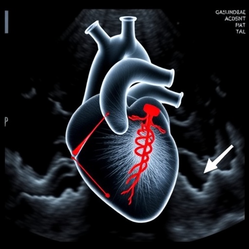
This ultrasound-guided technique utilizes high-resolution imaging to visualize cardiac structures during injection, allowing for precise needle placement into the left ventricle. By honing in on the left heart region, ULI leverages immediate proximity to the ascending thoracic aorta, facilitating efficient vector dispersal directly to the target tissue with limited systemic spillover.
Importantly, the adoption of ultrasound guidance transforms the injection procedure from a blind, high-risk intervention to a controlled, reproducible technique. This paradigm shift not only enhances animal welfare in preclinical studies but also strengthens the robustness of experimental outcomes by reducing variability commonly associated with injury or off-target effects.
In addition to improved safety and specificity, the ULI method exhibited increased transduction efficiency in the ascending thoracic aorta, which is particularly relevant given the high prevalence of aneurysms, dissections, and other genetic abnormalities in this region. Efficient gene delivery here could pave the way for targeted therapies that modify disease progression at the molecular level.
The ramifications of these findings extend beyond mouse models. The principles of ultrasound-guided injection could, in future, be adapted for larger animal studies and ultimately for human therapeutic interventions, imparting a clinical utility that overcomes current delivery barriers in aortic gene therapy.
Furthermore, by reducing hepatotoxicity associated with systemic vector administration, the ULI technique may enable higher vector doses or repeated administration, which are essential for robust and prolonged therapeutic gene expression in chronic aortic diseases. This opens new avenues for dosing strategies that were previously limited by safety concerns.
The study also highlights the critical role of cutting-edge imaging modalities in advancing gene therapy protocols. Ultrasound imaging is relatively accessible, non-invasive, and cost-effective, making it an attractive tool for widespread adoption in preclinical and possibly clinical gene delivery procedures.
Given the complexity in translating gene therapy from bench to bedside, methodologies like ULI that harmonize efficiency with safety represent vital steps toward viable treatments. Diseases such as Marfan syndrome, Loeys-Dietz syndrome, and other inherited thoracic aortic conditions could particularly benefit from gene therapies enabled by such innovative injection approaches.
While the current work focuses on technical delivery aspects, it sets the stage for subsequent studies exploring therapeutic efficacy, long-term expression profiles, and the immune response following ULI-mediated gene transfer. These investigations will be crucial to validate this method’s full potential in treating thoracic aortic pathologies.
In sum, the introduction of ultrasound-guided left heart injection marks a significant milestone in the quest for precision gene therapy targeting the thoracic aorta. By marrying real-time imaging with refined injection techniques, researchers have devised a method that optimizes transduction efficiency while minimizing procedural risks and off-target effects.
As such, this advancement holds promise for accelerating preclinical research and enhancing the translatability of gene therapy approaches aimed at this life-threatening vascular disease. With further refinement and adaptation, ULI may become a cornerstone technique in the future armamentarium against genetic aortic disorders.
The findings of this study underscore the transformative potential of integrating imaging technology into gene delivery protocols and exemplify the innovative spirit driving the next generation of cardiovascular therapeutics. The fusion of molecular biology, imaging, and interventional technique marks a new frontier in precision medicine.
Future research will undoubtedly delve deeper into the mechanistic underpinnings of ULI-enhanced gene transduction and explore its applicability across other cardiovascular targets. The journey from these insights to clinical breakthroughs is poised to redefine how we conquer complex genetic diseases of the aorta.
Subject of Research: Gene delivery techniques for the thoracic aorta in murine models focusing on safety and efficiency improvements using ultrasound guidance.
Article Title: Ultrasound-guided left heart injection: a safer and more efficient strategy for mouse thoracic aortic gene delivery.
Article References:
Yang, Y., Xu, K., Yuan, Y. et al. Ultrasound-guided left heart injection: a safer and more efficient strategy for mouse thoracic aortic gene delivery. Gene Ther (2026). https://doi.org/10.1038/s41434-026-00603-7
Image Credits: AI Generated
DOI: 10.1038/s41434-026-00603-7
Keywords: thoracic aortic disease, gene therapy, adeno-associated virus, ultrasound-guided injection, gene delivery, murine model, hepatotoxicity, cardiovascular genetics
Tags: cardiovascular gene therapy advancementshepatotoxicity in gene therapyimproving gene therapy safetyleft heart injection techniquemurine models for gene deliverypercutaneous cardiac injection riskspreclinical cardiovascular researchreducing off-target gene therapy effectssafer gene delivery methodstargeted aortic gene therapythoracic aortic disease treatmentultrasound-guided gene delivery


