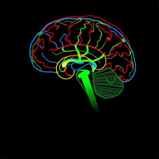
In a groundbreaking study published in the prestigious journal Nature, researchers at UCLA have unraveled some of the intricate neural circuits within the mouse medial prefrontal cortex (mPFC) that orchestrate the brain’s response to stress and social behavior. This milestone in neuroscience not only advances our understanding of fundamental brain processes but also paves the way for innovative treatments targeting complex neuropsychiatric disorders such as post-traumatic stress disorder (PTSD), depression, and anxiety.
The medial prefrontal cortex, a critical region nestled in the frontal lobes of the brain, has long been recognized for its role in personality, decision-making, and emotional regulation. Despite decades of research, the exact circuitry by which this brain region integrates myriad sensory inputs with internal physiological states to produce adaptive or maladaptive behavioral responses remained elusive. The UCLA team employed cutting-edge techniques—combining genetic labeling strategies, high-resolution 3D brain imaging, and artificial intelligence-driven circuit mapping—to dissect the fine-scale connectivity and organization of mouse mPFC subregions, specifically the dorsal peduncular area (DP) and infralimbic area (ILA).
Employing genetically encoded tracers, the researchers traced neuronal projections and synaptic partners within the mPFC, constructing a detailed wiring diagram of these visceromotor hubs. These areas act as integrative nodes, synthesizing information related to external sensory stimuli and internal bodily signals, such as those from the autonomic nervous system, to coordinate behavioral and physiological responses to stress. The sophisticated AI tools developed for this study enabled the automated reconstruction of neuronal circuits in three dimensions, revealing previously unseen patterns of connectivity and interregional communication.
One of the most compelling insights from this work concerns how these mPFC hubs not only modulate emotional reactivity but maintain emotional stability through balanced excitatory and inhibitory circuits. Dysregulations in this delicate balance could underlie the emotional instability observed in myriad psychiatric disorders. By elucidating the precise synaptic arrangements and molecular identities of these neurons, the study provides a cellular-level blueprint that parallels similar visceromotor circuits conserved in the human ventromedial prefrontal cortex (vmPFC).
The implications of these findings echo a historical neuroscience narrative dating back over 170 years to the famous case of Phineas Gage, a railroad worker who survived a traumatic frontal lobe injury yet underwent profound personality changes. Gage’s case underscored the significance of the prefrontal cortex in governing social behavior and emotional regulation. However, the neural underpinnings of such personality alterations have long remained a mystery. This research takes a pivotal step toward filling that knowledge gap and directly links mPFC circuitry to the regulation of complex behaviors and stress responses.
Moreover, the study’s integration of advanced 3D reconstructions with AI-driven analysis sets a new standard for investigating brain architecture at the mesoscale level. This methodological breakthrough not only accelerates data acquisition and analysis but also enhances reproducibility, offering an unprecedented resolution for mapping brain circuits involved in neuropsychiatric disorder pathophysiology.
Beyond the fundamental scientific advancements, this work holds profound clinical relevance. By pinpointing the neuronal circuits that orchestrate physiological and emotional responses to stress, the UCLA team offers promising targets for the development of novel, precision-based therapeutic interventions. These may one day include targeted neuromodulation, pharmacological agents aimed at circuit-specific molecular markers, and improved diagnostic tools capable of identifying early signs of neuropsychiatric dysfunction.
Additionally, the study raises intriguing questions about the interaction between brain regions responsible for integrating internal bodily states and those processing external environmental information. Understanding how these networks synchronize to generate coherent behavior under stress has enormous implications for tackling disorders characterized by impaired emotional regulation and social cognition.
The researchers underscore that the cellular and circuit-level insights gained from mice are highly relevant to human brain function due to evolutionary conservation of mPFC structures and connectivity patterns. This conservation bolsters the translational potential of the findings, suggesting that future therapies targeting homologous human brain circuits could mitigate the debilitating effects of mood and anxiety disorders.
Furthermore, this research exemplifies how multidisciplinary approaches—bridging genetics, neuroanatomy, computational modeling, and behavioral neuroscience—can unravel the complexities of brain function. Such holistic perspectives are vital for deciphering the labyrinth of neural interactions that underlie human cognition, emotion, and behavior.
In summary, the UCLA-led study provides a seminal contribution to neuroscience by delivering a comprehensive, high-resolution map of the mouse medial prefrontal cortex’s visceromotor circuits. This work not only enriches our fundamental understanding of emotional and stress regulation but also charts a course toward innovative interventions addressing some of the most pressing challenges in mental health today. As Dr. Hong Wei Dong, the study’s lead author and director of the UCLA Brain Research & Artificial Intelligence Nexus, eloquently stated, this is “a wiring diagram of one of the brain’s most mysterious control centers,” opening the floodgates to targeted therapies for stress-related and social dysfunction disorders.
Subject of Research: Animals
Article Title: Neural networks of the mouse visceromotor cortex
News Publication Date: 27-Aug-2025
Web References: https://www.nature.com/articles/s41586-025-09360-w, http://dx.doi.org/10.1038/s41586-025-09360-w
References: Dong, H.W., et al. (2025). Neural networks of the mouse visceromotor cortex. Nature. DOI: 10.1038/s41586-025-09360-w
Keywords: Behavioral neuroscience, Psychiatry, Mental health, Psychiatric disorders, Neuroscience, Anxiety disorders, Behavior disorders
Tags: artificial intelligence in brain mappingbrain circuits regulating stressemotional regulation mechanismsgenetic labeling in neurosciencehigh-resolution brain imaging techniquesmedial prefrontal cortex functionsneuronal connectivity in mPFCneuropsychiatric disorder treatmentsPTSD and anxiety researchsocial behavior in micesynaptic organization in brain regionsUCLA neuroscience research


