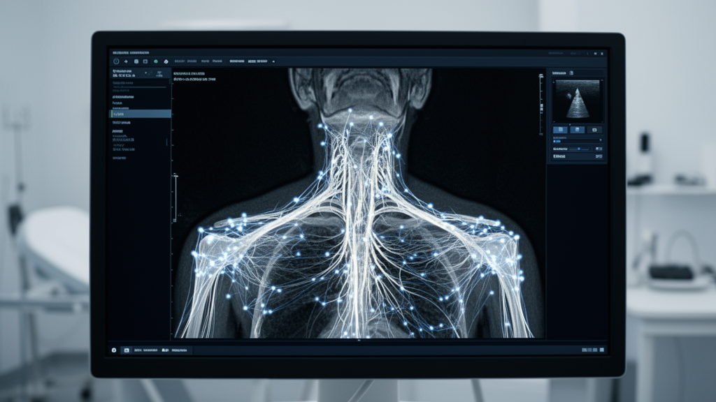
A deep learning architecture combining transformer-based semantic reasoning and spatial edge enhancement has achieved an average precision of 0.407 for nerve detection and an overall mean average precision of 0.583 in ultrasound images of the brachial plexus. Developed to address the severe acoustic degradation inherent to ultrasound guidance, the framework—designated as transformer-based detection with semantic and spatial feature fusion, or TDSS—introduces three plug-and-play modules designed to resolve anatomical ambiguities during peripheral nerve block procedures. Experimental evaluations conducted on the dedicated Ultrasound Brachial Plexus Dataset demonstrated that the system consistently outperformed established convolutional and vision transformer models across multiple anatomical structures. By integrating edge-sensitive structural priors, cross-scale attention, and self-distillation mechanisms into an end-to-end detection pipeline, the architecture provides verifiable localization gains for subtle neural targets that are frequently obscured by acoustic interference.
Peripheral nerve blocks represent a vital clinical technique in modern surgical anesthesia, wherein clinicians inject local anesthetics into tissue surrounding targeted nerve pathways to induce regional anesthesia, decrease postoperative pain, shorten hospital recovery intervals, and improve patient comfort. Under standard clinical workflows, ultrasound guidance enables anesthesiologists to identify essential neuroanatomical structures—specifically nerves, arteries, veins, and surrounding muscular layers—while tracking needle trajectories and the dispersion of injected anesthetics. However, acoustic positioning of the brachial plexus and its adjacent vascular boundaries currently depends on manual visual interpretation by experienced medical personnel, a procedure that remains time-intensive and heavily reliant on specialized clinical expertise. Ultrasound imaging inherently exhibits heavy speckle noise, low spatial resolution, and minimal acoustic impedance variation between neural structures and surrounding anatomical backgrounds. These physical constraints obscure semantic boundaries, complicate clinical delineation, and hinder the manual creation of large-scale annotated medical training repositories.
To overcome these imaging hurdles, the TDSS network operates as an integrated end-to-end framework organized around a standard convolutional backbone coupled to a transformer encoder-decoder structure and three modular components: the spatial edge adaptive enhancement module, the multi-scale semantic fusion module, and the spatial distillation-assisted detection module. In this structural workflow, input ultrasound frames are first processed by the spatial edge adaptive enhancement component to generate edge-aware structural representations before passing through a ResNet-50 feature extractor. The extracted feature hierarchy is subsequently structured into a multi-resolution feature pyramid within the multi-scale semantic fusion module, which applies channel and spatial cross-attention to resolve dependencies across scales. Global semantic features are further synthesized within the transformer encoder via self-attention mechanisms, while the transformer decoder and dedicated detection heads generate categorical classifications and bounding box coordinates, guided concurrently by spatial distillation to refine structural localization.
The spatial edge adaptive enhancement module functions as a low-level structural prior that emulates the biological visual system’s heightened sensitivity to physical edges while discarding background noise. For an input image, the module applies a Canny gradient operator utilizing lower and upper thresholds of 40 and 120 to extract a directional edge map. Rather than treating this edge extraction as a final anatomical contour, the system concatenates the edge map with the original input image to form a composite representation. This composite input is subsequently processed through a sequence of convolutional network blocks comprising 3×3 convolution kernels, batch normalization layers, and rectified linear unit activations. Merging these processed edge maps with hybrid convolutional features generates an intermediate feature map that passes through a sigmoid activation function to construct an attention map. Computing the inner product of this attention distribution with the mapped features produces a refined spatial edge map, suppressing acoustic speckle artifacts before deep feature fusion.
Addressing the wide structural diversity across tissue categories, the multi-scale semantic fusion module incorporates a cross-scale attention mechanism to replace simple feature pyramid interactions. Because anatomical structures within ultrasound scans range from compact nerve bundles to extensive muscle groups, the module uses multi-scale patch embeddings inspired by the Swin transformer. For each feature map across the pyramid, patch dimensions are scaled progressively, and tokens are extracted via average pooling followed by 1×1 depthwise separable convolutions that project each scale to an equivalent token representation. Tokens from multiple levels undergo layer normalization and concatenation along the channel dimension to establish linked queries and keys, while individual scale tokens provide values. Scaled dot-product attention computes spatial dependencies across the concatenated representations, producing multi-scale attention maps. The output passes through depthwise convolutions, layer normalization, and Gaussian error linear unit activations to generate synthesized features that capture both fine structural boundaries and global contextual relationships.
To resolve bounding box drift caused by ambiguous ultrasound boundaries, the spatial distillation-assisted detection module applies self-distillation and multiple instance learning to refine target localization. Recognizing that spatial localization in ultrasound presents greater computational difficulty than categorical classification, the module constructs perturbed proposal packages around candidate bounding boxes. Each proposal package contains eight candidate proposals generated by perturbing scale, aspect ratio, and center offsets within defined bounds, specifically applying a scale-jitter range of 0.20 and a center-jitter range of 0.10. RoI Align and shared fully connected layers extract features from these packages, while classification scores and spatial consistency constraints—derived from predicted intersection over union values and center distances—are evaluated to select the top three refined proposals. During model training, the framework optimizes an integrated loss function balancing standard detection losses, which combine L1, generalized intersection over union, and focal losses, with distillation loss weighted by a hyperparameter of 0.35.
The quantitative evaluation of TDSS was performed on the Ultrasound Brachial Plexus Dataset, a specialized medical imaging benchmark comprising 2035 annotated ultrasound frames collected from 200 human patients. Imaging data were acquired using two distinct clinical systems—the Siemens ACUSON NX3 Elite and the Philips EPIQ5—to incorporate hardware-level variation into the benchmark. Experienced anesthesiologists annotated all frames, identifying four target anatomical classes: arteries, veins, nerves, and muscle tissue. To ensure rigorous evaluation without data leakage, the investigators partitioned the data at the patient level, allocating 1835 images from 180 patients to the training set and 200 images from 20 patients to the held-out test set. Measurement of object-size distributions within the benchmark confirmed significant geometric disparity across anatomical structures: nerve instances were dominated by small and medium pixel areas with subtle boundaries, whereas muscular regions comprised large, distributed contours.
Experimental implementation of the TDSS architecture was executed in PyTorch on an NVIDIA A100 graphics processing unit utilizing mixed-precision computation across a 24-epoch training schedule. All input ultrasound images were standardized to dimensions of 640 by 640 pixels using bilinear interpolation. The training process utilized the AdamW optimizer with a weight decay parameter of 0.0001, an initial learning rate of 0.01, and a batch size of 4, reducing the learning rate by a factor of ten at epoch 16 and again at epoch 22. To prevent overfitting and enhance resistance to acoustic disturbances, the researchers incorporated data augmentation pipelines including random horizontal flipping with a 0.5 probability, random scaling between 0.8 and 1.2, boundary-clearing cropping, and Gaussian noise injection. Controlled comparisons maintained identical patient-level data partitions, input dimensions, and evaluation metrics across all tested models, using Mask R-CNN with ResNet-50 and a feature pyramid network as the primary baseline.
On the test set of the Ultrasound Brachial Plexus Dataset, TDSS achieved superior precision across all evaluated categories when compared with established detection baselines. For nerve detection, TDSS reached an average precision of 0.407, surpassing DINO at 0.392, RTMDet at 0.390, Grounding DINO at 0.385, and YOLOv9 at 0.380. Under the stricter localization criterion of average precision at an intersection over union threshold of 0.75, TDSS achieved a score of 0.593, outperforming DINO at 0.583, RTMDet at 0.585, Grounding DINO at 0.580, and YOLOv9 at 0.575. Evaluation of the foundation segmentation model MedSAM on the benchmark yielded suboptimal results, which the researchers attributed to a substantial domain gap between natural pretraining datasets and the specialized acoustic characteristics of brachial plexus ultrasound scans. The performance advantage of TDSS at elevated intersection over union thresholds demonstrated that its edge-aware and distillation mechanisms substantially improved high-precision boundary localization for clinically critical neural structures.
Component-level ablation experiments and modular plug-and-play evaluations confirmed the individual and cumulative contributions of each introduced architectural element. When individually inserted into a baseline Mask R-CNN framework with a default mean average precision of 0.395, the spatial edge adaptive enhancement module increased the overall score to 0.414, delivering an absolute gain of 1.9 percentage points. The multi-scale semantic fusion module independently raised the baseline score to 0.416, representing an absolute improvement of 2.1 percentage points, while the spatial distillation-assisted detection module improved baseline performance to 0.402, an increase of 0.7 percentage points. In cumulative ablation evaluations within the full TDSS pipeline, the combined modules achieved category-specific average precision scores of 0.712 for arteries, 0.691 for veins, 0.407 for nerves, and 0.531 for muscle tissues. Future research directions outlined by the authors include evaluating cross-modality generalizability across alternate anatomical regions, developing unsupervised annotation strategies, and integrating the framework into intraoperative guidance hardware.
Subject of Research: Deep learning multi-object detection in brachial plexus ultrasound imaging
Article Title: Enhanced multi-object detection in brachial plexus ultrasound images using transformer-based semantic and spatial feature fusion
Article References: Wei, H., Gao, X., Zhong, Y., Ding, Y., & Han, T. (2026). Enhanced multi-object detection in brachial plexus ultrasound images using transformer-based semantic and spatial feature fusion. Visual Computing for Industry, Biomedicine, and Art, 9(1), Article 27. https://doi.org/10.1186/s42492-026-00236-y
Image Credits: AI Generated
DOI: 10.1186/s42492-026-00236-y
Keywords: Brachial Plexus, Ultrasound Imaging, Deep Learning, Object Detection, Transformer, Peripheral Nerve Block, TDSS, Semantic Feature Fusion, Spatial Edge Adaptive Enhancement, Multi-Scale Semantic Fusion, Spatial Distillation, Computer Vision, Medical Image Analysis, Image Segmentation, Medical AI, Acoustic Speckle Noise, Neural Structures
News Source: Blake Davidson. (November 22, 2026). Transformer Framework Enhances Multi-Object Detection in Brachial Plexus Ultrasound Imaging. Scienmag.