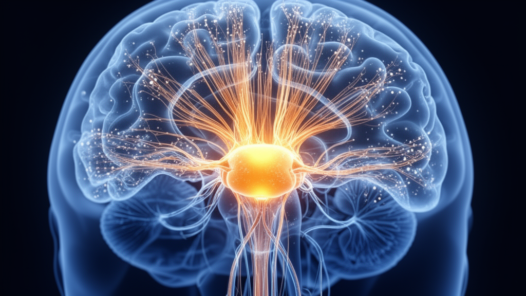
Researchers at the University of Electronic Science and Technology of China have identified a disruption in the functional hierarchy of the thalamus in patients with idiopathic generalized epilepsy (IGE). Published in European Radiology Experimental, the study utilized resting-state functional magnetic resonance imaging (fMRI) to map how thalamic connectivity varies across different strength levels. The findings suggest that the thalamus fails to maintain its typical organizational structure, a defect that correlates with poorer working memory performance in affected individuals.
The thalamus acts as a central hub in the brain, synchronizing oscillations and relaying signals between the cerebral cortex and the cerebellum. In IGE, a condition characterized by generalized tonic-clonic seizures and 2.5 to 5 Hz spike-wave discharges, the thalamo-cortical network is a core pathological substrate. While previous studies have noted thalamic hyperactivation during seizures, the specific impact of epilepsy on the thalamus’s hierarchical organization remained poorly understood until now.
To investigate this, the study team collected fMRI data from 144 IGE patients and 222 healthy controls. The participants were recruited from the neurology department of the Affiliated Hospital of the University of Electronic Science and Technology of China. Diagnosis for the IGE cohort was confirmed by two independent neurologists according to International League Against Epilepsy criteria. The healthy control group was demographically matched to the patient group in terms of age and sex.
The analytical approach involved computing voxel-wise functional connectivity between the thalamus and both the cerebrum and the cerebellum. Rather than analyzing connectivity as a single uniform metric, the researchers divided the connection strengths into ten percentile-based bins. This allowed them to examine how the thalamic hierarchy changes across varying connectivity strengths, from strongly anticorrelated to strongly correlated connections. This method helps capture the stability or reorganization of hierarchical organization across different coupling conditions.
In healthy controls, the thalamus exhibited a clear connectome-dependent reorganization. Under anticorrelated conditions, the thalamus predominantly coupled with low-order unimodal networks, such as sensorimotor and visual regions. In contrast, under correlated conditions, it coupled with high-order cognitive networks. This pattern reflects the thalamus’s role as an integrative hub, balancing segregation and integration for hierarchical information transfer between sensory and cognitive processing.
In patients with IGE, this hierarchical organization was significantly disrupted. The study found that IGE patients showed altered functional connectivity profiles in the limbic network, attentional network, and cerebellar systems. Specifically, under anticorrelated conditions, IGE patients exhibited higher proportions of connections within the dorsal and ventral attention networks compared to healthy controls. Conversely, under correlated conditions, these attentional network proportions were reduced, while the limbic network proportion increased. This suggests a failure of the thalamus to flexibly regulate interactions across different functional systems.
Further analysis using connectome gradient mapping revealed that the thalamic gradients in IGE patients showed marked distributional abnormalities across a broad range of connectivity strengths. The principal gradient in IGE patients followed different axes depending on the connectivity strength, shifting from a dorsomedial/ventrolateral axis to a dorsoanterior/ventroposterior axis at intermediate ranges. These gradient alterations indicate a non-uniform reorganization of thalamic functional hierarchies, potentially reflecting increased segregation and reduced cross-hierarchical integration.
The researchers also investigated thalamic eccentricity, a metric that indexes regional specialization within the gradient space. They found shared increases and decreases in eccentricity within motor and cognitive thalamic nuclei in IGE patients. Notably, specific clusters in the right center median nucleus and medial pulvinar showed decreased eccentricity, suggesting reduced segregation. Meanwhile, the left lateral posterior nucleus and medial pulvinar showed increased eccentricity, indicating enhanced differentiation but reduced cross-level integration.
The study found that thalamic eccentricity abnormalities were associated with poorer working memory performance in IGE patients. Specifically, two thalamo-cerebral clusters showing significant intergroup differences were positively associated with reaction time during an n-back working memory task. This association was observed in regions primarily located in the left lateral posterior nucleus, medial pulvinar, and lateral geniculate nucleus posterior part.
The study also examined the thalamo-cerebellar pathway, finding that decreased eccentricity in specific thalamic nuclei, including the mediodorsal nucleus parvocellular part and central lateral nucleus, was negatively correlated with reaction time. This suggests that disruption of the cerebello-thalamic loop may underlie slower information processing and working memory deficits in IGE. The central lateral nucleus is a primary thalamic target for the cerebellum, and its dysfunction is known to impair motor coordination and arousal regulation.
The researchers conducted sensitivity and robustness analyses to ensure the reliability of their findings. They repeated the analyses using weighted instead of binarized connectivity matrices and with and without global signal regression. The results showed that the FC profiles and eccentricity findings were highly consistent across these different preprocessing strategies. However, the functional gradients showed some variable sensitivity to global signal regression, particularly in the third gradient under specific connectivity bins.
The study has several limitations. The cross-sectional design precludes determining whether the gradient alterations are a cause, consequence, or compensation of epilepsy. Additionally, the relatively low spatial resolution of the fMRI data may limit the spatial precision of the results. Working memory performance was not assessed in the healthy control group, although comparisons with an independent matched sample showed significantly lower accuracy and longer reaction times in IGE patients. Antiseizure medication effects could also potentially influence the results, although additional analyses controlling for medication status yielded largely consistent findings.
These findings provide novel insights into the thalamic functional abnormalities in idiopathic generalized epilepsy. By demonstrating that the thalamic hierarchy is disrupted in a nucleus-specific manner, the study offers a systems-level explanation for the motor and cognitive deficits commonly observed in IGE. The results highlight the importance of examining connectivity across varying strengths to fully understand the complex network dysfunction associated with epilepsy.
Subject of Research: Disrupted thalamic functional hierarchy on resting-state fMRI in idiopathic generalized epilepsy
Article Title: Disrupted thalamic functional hierarchy on resting-state fMRI in idiopathic generalized epilepsy
Article References: Chen, J., Sun, X., Yu, K., Yang, Z., Li, H., Yang, Z., Chen, Y., Li, J., Rodriguez-Labrada, R., Yao, D., Jiang, S., & Luo, C. (2026). Disrupted thalamic functional hierarchy on resting-state fMRI in idiopathic generalized epilepsy. European Radiology Experimental, 10(1), Article 131. https://doi.org/10.1186/s41747-026-00788-x
Image Credits: AI Generated
DOI: 10.1186/s41747-026-00788-x
Keywords: Disrupted, thalamic, functional, hierarchy, resting-state, fMRI, idiopathic, generalized, epilepsy, scientific research
News Source: Kendall Mcintyre. (November 10, 2026). Thalamic Hierarchy Disrupted in Idiopathic Generalized Epilepsy. Scienmag.