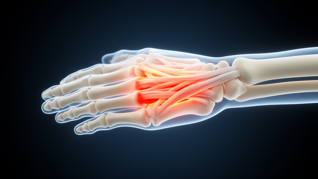
Imaging-detected tenosynovitis may be one of the earliest pathological events in inflammatory arthritis, according to a narrative review published in Advances in Rheumatology. The review, authored by Nicole Pamplona Bueno de Andrade, Rafael Mendonça da Silva Chakr and Claiton Viegas Brenol, brings together evidence on the role of tenosynovitis—inflammation of tendon sheaths—in the diagnosis and ongoing management of rheumatoid arthritis (RA).
The authors report that the histopathological features of tenosynovitis are indistinguishable from those of joint synovitis, the inflammation of the joint lining that is central to RA. Two modalities dominate its detection: musculoskeletal ultrasound (MSUS) and magnetic resonance imaging (MRI). According to the review, MSUS demonstrates high specificity, particularly when findings are compared with healthy controls or with individuals who do not go on to develop RA, whereas MRI generally offers superior sensitivity for identifying early inflammatory changes.
A central finding of the review is the predictive value of tenosynovitis. The authors state that it is a strong predictor of RA development, with studies highlighting an independent association between imaging-detected tenosynovitis and progression to RA. Beyond prediction, they note that tenosynovitis can enhance the diagnostic performance of established classification criteria, suggesting that adding imaging findings to existing frameworks may improve how early disease is identified.
The review also links tenosynovitis to disease activity. Imaging-detected tenosynovitis is correlated with clinical and laboratory parameters of disease activity, and it is described as highly responsive following disease-modifying antirheumatic drug (DMARD) therapy. This responsiveness, the authors argue, makes the finding valuable for monitoring disease activity and has led to its incorporation into various ultrasound scoring systems used in research and clinical practice.
Prognostically, the review reports that imaging-detected tenosynovitis is evident in predicting disease flares and radiographic progression in RA patients, meaning its presence on scans may signal a higher risk of worsening disease over time. The authors also point to emerging, though they emphasise limited, evidence suggesting a potential role in predicting treatment response—an area they identify as still developing rather than established.
As a narrative review, the article synthesises existing literature rather than presenting new patient data, and its conclusions reflect the state of published research on imaging-detected tenosynovitis in RA. For clinicians and researchers, the review’s message is that inflammation of tendon sheaths, when detected on imaging, carries information that extends well beyond the immediate scan—informing who may progress to RA, how active disease is, and how patients may fare over time.
Subject of Research: The role of imaging-detected tenosynovitis in the diagnosis and monitoring of rheumatoid arthritis
Article Title: Imaging-detected tenosynovitis in rheumatoid arthritis: from early diagnosis to disease monitoring
Article References: de Andrade, N. P. B., da Silva Chakr, R. M., & Brenol, C. V. (2026). Imaging-detected tenosynovitis in rheumatoid arthritis: from early diagnosis to disease monitoring. Advances in Rheumatology. https://doi.org/10.1186/s42358-026-00556-w
Image Credits: AI Generated
DOI: 10.1186/s42358-026-00556-w
Keywords: rheumatoid arthritis, tenosynovitis, musculoskeletal ultrasound, MRI, early diagnosis, disease monitoring, DMARD therapy, disease activity, prognosis, narrative review, Advances in Rheumatology, Imaging-detected
News Source: Ophelia Keating. (October 29, 2026). Tenosynovitis on imaging emerges as early warning sign of rheumatoid arthritis. Scienmag.