Chronic recurrent temporomandibular joint (TMJ) dislocation presents a complex therapeutic challenge characterized by debilitating functional impairment, pain, and the necessity for manual reduction of the condyle following anterior translation beyond the articular eminence. Surgical strategies traditionally employed to mitigate these symptoms vary widely, often relying on freehand bony modifications or eminence-blocking procedures that lack standardized, reproducible outcomes. A recent prospective study has evaluated a template-guided, digitally planned patient-specific implant augmentation eminoplasty as a potential solution to enhance the precision of these surgical interventions. By integrating high-resolution computed tomography (CT) data with computer-aided design and computer-aided manufacturing (CAD/CAM) technologies, this approach aims to provide a controlled mechanical stop to restrict excessive condylar movement while minimizing the inherent variability associated with intraoperative estimation.
The study cohort consisted of eleven patients, all older than sixteen years, who suffered from chronic recurrent TMJ dislocation or hypermobility, defined in this clinical research as a maximal incisal opening (MIO) exceeding 40 millimeters. Patients with specific comorbidities, including epilepsy, psychological disorders, or prior TMJ fractures, were excluded to maintain a relatively homogenous study population. Prior to surgical intervention, all participants underwent comprehensive clinical and radiologic evaluation, including multi-slice computed tomography (MSCT) and panoramic TMJ views in open and closed positions, to map the individual anatomy of the zygomatic arch and the articular eminence. This preoperative diagnostic framework ensured that the subsequent implant design was tailored to the unique patient anatomy, facilitating stable seating and optimized load distribution across the joint structures.
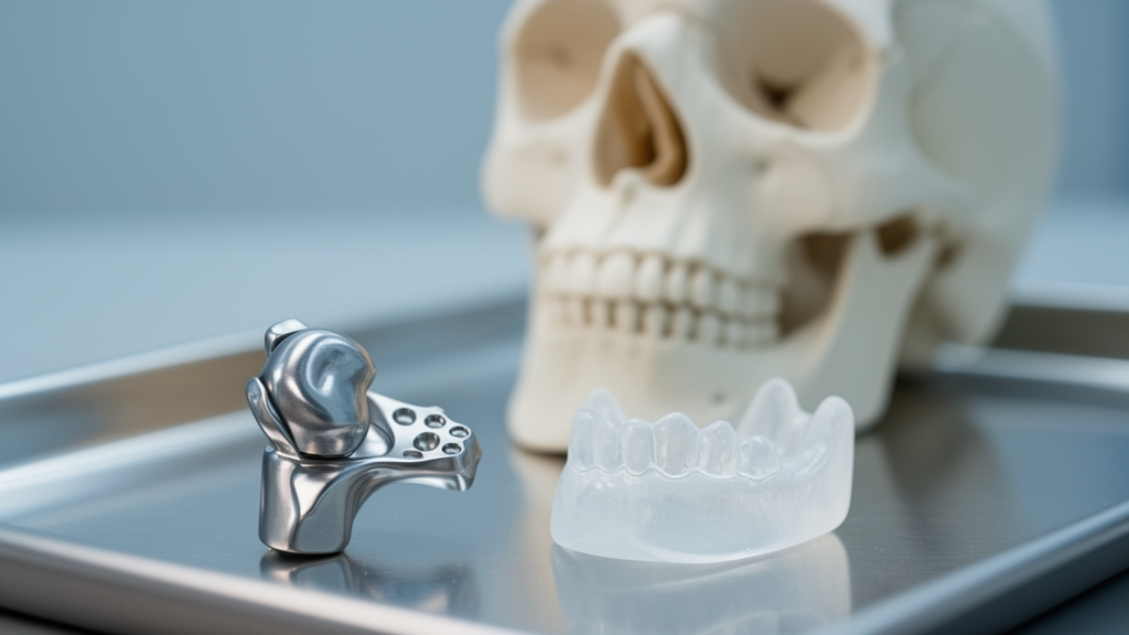
The procedural workflow involved a sophisticated digital pipeline beginning with the generation of a three-dimensional cranio-maxillofacial model in the Mimics Innovation Suite. Patient-specific implants were manufactured using ultrahigh molecular weight polyethylene (UHMWPE), a material widely recognized for its biocompatibility and favorable tribological properties in orthopedic applications. Concurrently, digital templates were fabricated through stereolithography (SLA) resin printing. These guides were meticulously designed to reference stable anatomic landmarks along the zygomatic arch, ensuring that the surgeon could consistently reproduce pre-planned drilling trajectories and screw-entry points. During the surgery, performed under general anesthesia, the guide allowed for the standardized placement of four titanium screws, securing the implant against the articular eminence to provide the desired mechanical reinforcement.
Clinical outcomes were assessed through visual analogue scale (VAS) pain scores and measurements of maximal interincisal opening at baseline, two weeks, and six months postoperatively. The results demonstrated a statistically significant reduction in pain across the cohort. The combined mean VAS pain score decreased from 8.80 ± 0.53 preoperatively to 8.07 ± 0.73 at the two-week assessment and further to 6.29 ± 1.29 at the six-month follow-up. Pairwise comparisons across these intervals were identified as highly significant, with p-values below 0.001. This reduction in pain is hypothesized to stem from a combination of successful physical stabilization, decreased soft-tissue inflammation over time, and increased patient confidence in functional jaw movements, although the multifactorial nature of TMJ pain requires cautious interpretation.
Parallel to the reduction in pain, the study measured a substantial decrease in maximal interincisal opening, a critical metric for evaluating the efficacy of the mechanical block created by the augmentation implant. The preoperative mean mouth opening of 45.68 ± 1.78 mm was successfully reduced intraoperatively to 32.55 ± 3.45 mm, further stabilizing to 30.64 ± 3.32 mm at two weeks and 28.73 ± 3.93 mm at the six-month mark. Statistical analysis indicated a significant decline from baseline levels, with no significant difference between the two-week and six-month time points (p = 0.082). This stability suggests that the surgical approach achieved its primary mechanical goal of restricting the extreme range of motion that previously predisposed the condyle to recurrent dislocation events.
Despite these clinical improvements, the study identified a concerning complication rate, with two patients (18.2%) developing postoperative infections on the right side. In both instances, clinical signs such as pain, swelling, and erythema emerged approximately three weeks after the procedure. Initial conservative management with antibiotics and cold compression provided only transient relief. Subsequent recurrence of symptoms and the development of purulent discharge by the two-month mark necessitated the removal of the implants. Microbiological data were not collected as the decision for explantation followed the emergence of visible discharge. However, it is notable that following the removal of these implants, none of these patients experienced a clinical recurrence of TMJ dislocation during the remaining follow-up period, suggesting that early stabilization or subsequent periarticular fibrosis might have contributed to joint control.
Radiologic assessments at the six-month milestone provided further insights into the long-term structural integrity of the technique. MSCT imaging did not reveal any evidence of implant malposition, osteolysis, or screw fractures in the patients who retained their implants. This reinforces the hypothesis that anatomically conforming, patient-specific prostheses offer a more predictable interface than constructs adapted intraoperatively. By minimizing micromotion at the bone-implant interface, this personalized geometry likely contributes to the observed stability. Nevertheless, the reliance on UHMWPE presents a limitation regarding radiologic monitoring, as its radiolucent nature necessitates indirect assessment of the implant’s position and stability within the confined preauricular surgical site.
From a biomechanical perspective, the introduction of an augmentation implant represents a divergence from procedures such as eminectomy, which seeks to remove structural barriers. By creating a mechanical stop, eminoplasty intentionally alters joint kinematics. While this effectively manages the primary complaint of instability, it poses potential long-term questions regarding load distribution across the disc, condyle, and glenoid fossa. Future research must determine whether these alterations in joint movement contribute to subclinical remodeling or degenerative changes over longer timelines. The study’s focus was intentionally limited to short-term functional and pain outcomes, and the absence of long-term longitudinal surveillance remains a significant gap in the current understanding of the procedure’s biomechanical impact.
The study’s conclusions are constrained by several limitations, including the small sample size, the absence of a comparator cohort, and the relatively short duration of follow-up. The exclusion of patients with pre-existing TMJ internal derangement further limits the generalizability of the findings to a broader clinical population where such degenerative pathologies are common. Furthermore, the absence of validated functional outcome instruments, such as the Jaw Functional Limitation Scale or the Oral Health Impact Profile, means the study could not comprehensively assess the patient-reported impact of the reduced mouth opening on activities of daily living, such as mastication, oral hygiene, and routine dental care.
As the field of maxillofacial surgery continues to evolve, the integration of digital planning into traditional techniques such as eminoplasty offers a pathway to higher precision. However, this preliminary case series emphasizes that technological advancements do not eliminate biological risks, particularly in the context of wound healing, bacterial contamination, and soft-tissue management. The observed infection rate highlights that even with superior implant seating and reduced mechanical variability, host factors and surgical handling remain pivotal determinants of procedural success. Future comparative studies are essential to delineate the role of this technique relative to existing, well-established surgical options such as eminectomy.
The selection of biomaterials such as UHMWPE, PEEK, and titanium should be considered an integral component of future surgical planning, as each material offers a unique balance of mechanical strength, radiolucency, infection risk, and manufacturing complexity. Establishing a standardized core outcome set that includes pain, functional limitation metrics, complication reporting, and long-term radiologic stability is critical for future research. Only through such rigorous, controlled evaluations can clinicians define the optimal postoperative mouth opening thresholds that successfully balance joint stability with the preservation of functional quality of life for patients suffering from chronic recurrent TMJ dislocation.
Subject of Research: Clinical outcomes and safety of template-guided patient-specific implant augmentation eminoplasty for chronic recurrent temporomandibular joint dislocation.
Article Title: Template-guided digitally planned patient-specific implant augmentation eminoplasty for chronic recurrent temporomandibular joint dislocation: a prospective study
Article References: Ibrahim, M. G., Swaify, G. A., Zaitoun, I., Medra, A. M., Kosba, A. Y., & Abdeldayem, M. (2026). Template-guided digitally planned patient-specific implant augmentation eminoplasty for chronic recurrent temporomandibular joint dislocation: a prospective study. Maxillofacial Plastic and Reconstructive Surgery, 48(1), Article 10. https://doi.org/10.1186/s40902-026-00508-w
Image Credits: AI Generated
DOI: 10.1186/s40902-026-00508-w
Keywords: Temporomandibular Joint Disorder, TMJ Dislocation, Eminoplasty, Patient-Specific Implants, Digital Planning, CAD/CAM Dentistry, UHMWPE, Surgical Navigation, Maxillofacial Surgery, Chronic Recurrent Dislocation, Mandibular Kinematics, Postoperative Infection, Clinical Outcomes, Orthopedic Biomaterials, Surgical Reproducibility
News Source: Ophelia Keating. (November 18, 2026). Template-Guided Patient-Specific Implants Reduce Chronic Temporomandibular Joint Dislocation Symptoms. Scienmag.