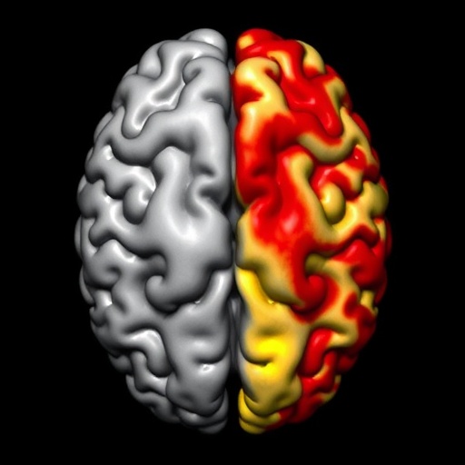
In a groundbreaking new study published in Nature Communications, researchers have unveiled compelling evidence linking the asymmetric accumulation of two hallmark proteins in Alzheimer’s disease—tau and amyloid-beta—across cerebral hemispheres. This discovery sheds novel light on the spatial dynamics of the neurodegenerative process and could signify a paradigm shift in understanding why Alzheimer’s symptoms often manifest asymmetrically in patients. Delving into the intricate relationship between tau and amyloid pathology, the study opens up potential pathways for more targeted diagnostic and therapeutic strategies.
Alzheimer’s disease, a devastating neurodegenerative disorder, is characterized by the progressive decline in cognitive function accompanied by the buildup of abnormal protein aggregates in the brain. For decades, two proteins—amyloid-beta and tau—have been recognized as central players. Amyloid-beta plaques accumulate extracellularly, while tau forms neurofibrillary tangles inside neurons. The spatial and temporal patterns of these aggregates have been topics of intense study, but this new research emphasizes that their distribution is not always symmetrical across the brain’s hemispheres, challenging earlier assumptions of a relatively uniform pathology.
Using advanced neuroimaging techniques coupled with postmortem histopathological analysis, the study meticulously quantified the regional burden of tau and amyloid deposition in Alzheimer’s patients. Intriguingly, the data revealed that tau pathology tends to show hemispheric asymmetry that aligns with an uneven distribution of amyloid plaques. This coupling hints at a possible causative or facilitatory relationship, where the asymmetry of amyloid deposition might drive or influence the lateralization of tau pathology. Such an insight offers a biological explanation for why patients sometimes experience lateralized symptoms, such as predominantly left- or right-hemisphere cognitive impairments.
The research team employed positron emission tomography (PET) imaging tracers specific for tau and amyloid-beta to obtain in vivo visualization of protein distribution. This allowed for longitudinal tracking and high-resolution mapping of pathological load. Additionally, immunohistochemical staining of brain tissue samples validated the imaging findings at a microscopic level. The synergy between imaging and postmortem analysis provided robust, multidimensional evidence that the asymmetry is not an artifact but a reproducible hallmark of Alzheimer’s pathology at the population level.
Further analysis indicated that the degree of hemispheric asymmetry in tau correlated positively with the asymmetry of amyloid burden. This spatial correlation was most pronounced in key regions implicated in Alzheimer’s-related cognitive decline, including the medial temporal lobe and the posterior cingulate cortex. These regions are crucial for memory processing and executive function, aligning with clinical observations where asymmetric cognitive deficits correspond with more significant pathology on the affected side.
The biological underpinnings driving this asymmetry are complex but may stem from localized vulnerabilities in neuronal circuits or differential clearance mechanisms within hemispheres. The study hypothesizes that early amyloid accumulation on one side may create a microenvironment conducive to tau propagation, possibly via transneuronal spread or disruption of proteostatic systems. Understanding these pathways at a molecular and cellular level will be critical for future therapeutic interventions aiming to halt or reverse tau spreading.
This hemispheric asymmetry has profound implications for diagnosis. Conventional methods often assume bilateral, symmetric involvement and may overlook subtler, unilateral pathology. Incorporating assessments of asymmetrical tau and amyloid deposition into clinical protocols could enhance early diagnosis, particularly in atypical cases. Moreover, it may help refine prognostic models by recognizing that lateralized pathology might predict a distinct disease trajectory or response to treatment.
From a therapeutic perspective, strategies that can specifically target and modulate asymmetric amyloid or tau pathology could revolutionize Alzheimer’s care. For example, antibody-based therapies aimed at clearing amyloid or tau could be optimized to address the dominant hemisphere first or personalized based on the asymmetry profile. Such tailored approaches could maximize efficacy and minimize side effects, marking a significant departure from the conventional “one-size-fits-all” methodology.
The findings also raise fascinating questions about the relationship between structural and functional hemispheric asymmetries in the healthy brain and the progression of Alzheimer’s disease. It’s well-established that many cognitive functions, such as language and spatial reasoning, are lateralized to one hemisphere. The study suggests that these inherent asymmetries might influence vulnerability to pathological protein deposition, potentially explaining why disease manifestations are often side-biased.
Moreover, the interdisciplinary nature of the research, bridging neuroimaging, neuropathology, and clinical neuropsychology, exemplifies the power of integrated approaches in tackling complex brain disorders. The combination of cutting-edge PET imaging tracers with detailed neuropathological validation sets a benchmark for future studies aiming to unravel the multifaceted landscape of Alzheimer’s pathology.
The implications of the study extend beyond Alzheimer’s disease alone. Asymmetric patterns of neurodegeneration have been observed in other disorders such as frontotemporal dementia and Parkinson’s disease. The methodologies and principles outlined here could be adapted to investigate these conditions, potentially uncovering shared mechanisms of hemispheric vulnerability and disease progression.
One especially provocative aspect of the study is the potential for asymmetry to serve as a biomarker for disease staging or treatment monitoring. Quantitative metrics derived from the degree of hemispheric imbalance could be developed into clinical tools that track progression more sensitively than global measures of protein burden. This would enable clinicians to detect subtle changes earlier and adjust therapeutic regimens dynamically.
The researchers emphasize that future work should focus on longitudinal studies to establish causality between asymmetric amyloid and tau deposition. Understanding whether amyloid asymmetry precedes tau lobar localization or vice versa is key to unraveling the sequence of pathological events. Such knowledge would profoundly influence the timing and targets of interventional strategies.
In conclusion, this compelling research reframes Alzheimer’s disease pathology through the lens of hemispheric asymmetry, coupling two of its most notorious protein hallmarks in a spatially and functionally meaningful way. This nuanced understanding opens new avenues for diagnosis, treatment, and ultimately, the quest to decipher the enigmatic processes driving neurodegeneration. As the field advances, embracing the brain’s natural asymmetries may unlock novel opportunities to combat Alzheimer’s more effectively than ever before.
Subject of Research: Hemispheric asymmetry in tau and amyloid-beta protein deposition in Alzheimer’s disease.
Article Title: Hemispheric asymmetry of tau pathology is related to asymmetric amyloid deposition in Alzheimer’s Disease.
Article References:
Anijärv, T.E., Ossenkoppele, R., Smith, R. et al. Hemispheric asymmetry of tau pathology is related to asymmetric amyloid deposition in Alzheimer’s Disease. Nat Commun 16, 8232 (2025). https://doi.org/10.1038/s41467-025-63564-2
Image Credits: AI Generated
Tags: Alzheimer’s diagnostic strategiesAlzheimer’s disease researchamyloid beta depositsasymmetric brain pathologybrain hemisphere imbalancecognitive decline in Alzheimer’sneurodegenerative disordersneuroimaging techniques in Alzheimer’spostmortem histopathological analysisspatial dynamics of tau and amyloidtargeted Alzheimer’s therapiestau protein accumulation


