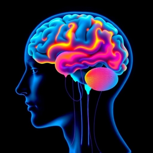

In a breakthrough study poised to reshape our understanding of Parkinson’s disease (PD), researchers have employed advanced synthetic magnetic resonance imaging (MRI) techniques to probe the intricate interplay between brain volume and subcortical myelin across different motor subtypes of the disease. Parkinson’s, a neurodegenerative disorder affecting millions worldwide, manifests heterogeneously, with diverse motor symptoms suggesting distinct underlying neuropathologies. This study spearheaded by Cheng, Wen, Ding, and colleagues, published in npj Parkinson’s Disease, elucidates how detailed imaging of brain microstructure can unravel these subtype-specific neurological variations, potentially steering us toward tailored therapeutic interventions.
Parkinson’s disease traditionally presents with hallmark motor symptoms such as tremor, rigidity, bradykinesia, and postural instability. However, these symptoms cluster into different motor subtypes — most notably tremor-dominant (TD), postural instability gait difficulty (PIGD), and intermediate forms — each hypothesized to involve unique neurodegenerative pathways. Precisely characterizing the cerebral underpinnings responsible for these clinical presentations has long remained a challenge in neuroscience, mainly due to limitations in conventional imaging modalities.
Central to this investigation is the use of synthetic MRI, a cutting-edge imaging modality that extends beyond mere anatomical visualization to quantitatively assess brain tissue properties. Unlike traditional MRI sequences that depend on distinct acquisitions, synthetic MRI generates multiple contrasting images via computational modeling from a single scan, vastly enhancing both efficiency and depth of tissue characterization. This enables researchers to measure parameters such as T1 and T2 relaxation times, proton density, and, importantly for this study, indicators reflecting myelin content and brain volume in subcortical regions — deep brain areas critically implicated in motor control.
.adsslot_1GkotVXFe2{width:728px !important;height:90px !important;}
@media(max-width:1199px){ .adsslot_1GkotVXFe2{width:468px !important;height:60px !important;}
}
@media(max-width:767px){ .adsslot_1GkotVXFe2{width:320px !important;height:50px !important;}
}
ADVERTISEMENT
The research team systematically analyzed a cohort of Parkinson’s patients classified into canonical motor subtypes, employing synthetic MRI to extract volumetric and myelin-related metrics from key subcortical structures like the substantia nigra, putamen, and thalamus. These regions are particularly relevant given their known involvement in dopaminergic signaling and motor circuit modulation, which are profoundly disrupted in Parkinson’s pathology. Sophisticated statistical models correlated the imaging findings with clinical assessments, revealing significant subtype-dependent discrepancies in both brain volume and subcortical myelin integrity.
One of the study’s pivotal revelations is the marked reduction in subcortical myelin observed predominantly within the PIGD motor subtype relative to the tremor-dominant group and healthy controls. This suggests that demyelination or myelin sheath degeneration may contribute more aggressively to gait and postural impairments than to tremor symptoms. Additionally, volumetric atrophy patterns diverged across subtypes, indicating that neurodegeneration in Parkinson’s is not monolithic but instead spatially and functionally heterogeneous. These nuanced insights challenge earlier conceptions of Parkinson’s as a uniform neurodegenerative entity.
The mechanistic implications of these findings are profound. Myelin, traditionally viewed in the context of white matter tracts, plays a crucial role in optimizing neural signal conduction velocity and synchrony across brain circuits. Subcortical myelin alterations could therefore disrupt key basal ganglia-thalamocortical loops responsible for motor execution, contributing to the phenotypic variations seen clinically. Furthermore, the observed volume reductions likely reflect combined neuronal loss, gliosis, and tract degeneration, which synergistically undermine motor function.
Integrating these imaging biomarkers with clinical phenotyping opens a new window onto the pathophysiology of Parkinson’s. The ability to non-invasively map myelin and volume changes in vivo provides a powerful tool for early diagnosis, prognostication, and monitoring disease progression. It may also illuminate the biological basis of differential treatment responses, such as the varying efficacy of dopaminergic therapies among subtypes, thereby guiding personalized medicine approaches.
Moreover, the application of synthetic MRI holds promise for enhancing longitudinal studies, as its quantitative nature reduces variability and allows for standardized cross-center comparisons. This is crucial in Parkinson’s research, where disease heterogeneity complicates the evaluation of novel therapeutics. Future clinical trials might integrate these imaging biomarkers as surrogate endpoints or inclusion criteria to refine patient stratification and maximize therapeutic impact.
Beyond its immediate clinical relevance, this study exemplifies the transformative potential of advanced imaging technologies in neuroscience research. By capturing multi-parametric data from a single scan, synthetic MRI marries efficiency with precision, overcoming the limitations of both traditional MRI and more laborious neuroimaging protocols. As computational models and scanner technologies further evolve, the depth and resolution of brain microstructural assessment will only improve, propelling discoveries in neurodegenerative diseases.
Equally compelling is the interdisciplinary collaboration harnessed in this work, spanning neurology, radiology, computational science, and biostatistics. Such a confluence is vital to decoding the complex biological signatures underpinning neurological disorders. This approach paves the way for integrative frameworks that combine imaging biomarkers with molecular, genetic, and clinical data to unravel Parkinson’s multifaceted nature.
While this research illuminates critical aspects of Parkinson’s neuropathology, it also raises intriguing questions. For instance, the temporal dynamics of myelin loss and volumetric atrophy across disease progression remain to be fully characterized. Does myelin degradation precede neuronal loss, or are these processes intertwined? Additionally, how do these imaging markers relate to non-motor symptoms often observed in Parkinson’s, such as cognitive impairment and autonomic dysfunction? Addressing these queries could further refine subtype definitions and inform comprehensive care strategies.
The potential for synthetic MRI extends beyond Parkinson’s disease. Its application could revolutionize the study of other neurodegenerative disorders marked by myelin pathology or structural brain changes, including multiple sclerosis, Alzheimer’s disease, and amyotrophic lateral sclerosis. Thus, this study not only advances Parkinson’s research but also showcases a versatile platform for neurological diagnostics more broadly.
In conclusion, Cheng and colleagues’ pioneering use of synthetic MRI reveals that subcortical myelin and brain volume alterations distinguish Parkinson’s motor subtypes, offering novel biomarkers with profound clinical implications. These insights deepen our comprehension of Parkinson’s heterogeneity and herald a new era where precision imaging informs personalized neuroscience. As synthetic MRI continues to mature, its integration into routine clinical and research settings could transform the landscape of neurodegenerative disease management, ultimately improving patient outcomes and quality of life.
Subject of Research:
Investigation of brain volume and subcortical myelin alterations in different motor subtypes of Parkinson’s disease using synthetic MRI.
Article Title:
Synthetic MRI study of brain volume and subcortical myelin in various Parkinson’s disease motor subtypes.
Article References:
Cheng, D., Wen, J., Ding, N. et al. Synthetic MRI study of brain volume and subcortical myelin in various Parkinson’s disease motor subtypes. npj Parkinsons Dis. 11, 255 (2025). https://doi.org/10.1038/s41531-025-01120-x
Image Credits: AI Generated
Tags: advanced imaging modalities in neurosciencebrain changes in Parkinson’s diseaseimaging brain microstructuremotor subtypes of Parkinson’sneurodegenerative disorder researchneurological variations in PDpostural instability gait difficultysubcortical myelin analysissynthetic MRI techniquestailored therapeutic interventions for PDtremor-dominant Parkinson’s subtypeunderstanding Parkinson’s disease symptoms

