A new study details how nutrient-starved cells divert protein transport stations to cellular recycling centers to be broken down, highlighting a novel approach cells use to deal with stressful conditions.
Credit: Liao et al.
A new study details how nutrient-starved cells divert protein transport stations to cellular recycling centers to be broken down, highlighting a novel approach cells use to deal with stressful conditions.
New proteins bound for outside the cell are manufactured on the endoplasmic reticulum (ER) – a snaking membrane inside the cell. Grape-like tubular outgrowths on the ER called ER exit sites serve as transport stations, collecting these newly synthesized proteins and delivering them to the next step in their journey.
In recent years, scientists have discovered that these ER exit sites also help deliver cellular material and misfolded proteins to lysosomes — organelles that degrade and recycle material in the cell — and provide a platform for replication of viruses, including COVID-19. But researchers were perplexed how this one structure, the ER exit site, can participate in all these diverse functions.
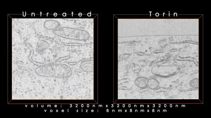
In a new study, researchers from HHMI’s Janelia Research Campus, led by Ya-Cheng Liao, a former postdoc in the Lippincott-Schwartz lab and now an assistant professor at Columbia University, used super-resolution live cell imaging and volume electron microscopy to examine the effect of nutrient stress on ER exit sites.
The team found that the stress triggers a series of molecules to work together to direct ER exit sites to lysosomes where they are destroyed – a novel pathway the cell may use to free up amino acids needed to make proteins inside the cell.
First, the researchers showed how the ER exit sites are delivered to and ingested by certain types of lysosomes when cells are starved of nutrients.
Next, the team detailed how this process happens. It starts when starving cells trigger the release of calcium from lysosomes. This causes an enzyme, ALG2, to get recruited to the ER exit sites where it binds to a structure called COPII that is attached to the neck that connects the ER to the ER exit site.
This connection between ALG2 and COPII starts a process called ubiquitination, which is involved in protein degradation. A protein on the lysosome involved in bringing cellular material to the organelle for destruction recognizes the ubiquitin produced by the ubiquitination process, driving the ER exit site to the lysosome.
Once at the lysosome, ALG2, which is attached to the ER exit site on one side, binds its other side to another protein, ALIX. ALIX interacts with ESCRT, a protein complex on the lysosome surface involved in ingestion. This interaction causes the ER exit site and the lysosome to become closer and closer until the ER exit site is engulfed and ingested by the lysosome.
Along with looking at this process in live cells, the team also reconstituted it in an artificial system, confirming how all the different components work together.
The new work details a novel pathway that cells use to combat stress, insight that could help researchers better understand how cells and organisms age. It could also shed light on other processes that involve the ER exit sites, including an unconventional way that viruses are delivered outside the cell through lysosomes, which could help researchers develop new treatments.
Journal
Developmental Cell
DOI
10.1016/j.devcel.2024.03.027
Article Title
COPII with ALG2 and ESCRTs control lysosome-dependent microautophagy of ER exit sites

