Credit: L.A. Cicero
Cells within our bodies divide and change over time, with thousands of chemical reactions occurring within each cell daily. This makes it difficult for scientists to understand what's happening inside. Now, tiny nanostraws developed by Stanford researchers offer a method of sampling cell contents without disrupting its natural processes.
A problem with the current method of cell sampling, called lysing, is that it ruptures the cell. Once the cell is destroyed, it can't be sampled from again. This new sampling system relies on tiny tubes 600 times smaller than a strand of hair that allow researchers to sample a single cell at a time. The nanostraws penetrate a cell's outer membrane, without damaging it, and draw out proteins and genetic material from the cell's salty interior.
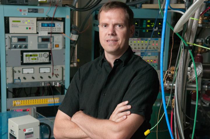
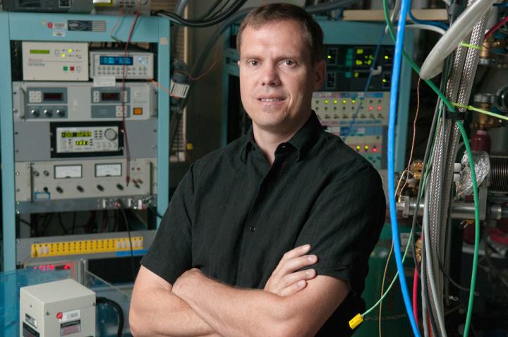
"It's like a blood draw for the cell," said Nicholas Melosh, an associate professor of materials science and engineering and senior author on a paper describing the work published recently in Proceedings of the National Academy of Sciences.
Non-destructive monitoring
The nanostraw sampling technique, according to Melosh, will significantly impact our understanding of cell development and could lead to much safer and effective medical therapies because the technique allows for long term, non-destructive monitoring.
"What we hope to do, using this technology, is to watch as these cells change over time and be able to infer how different environmental conditions and 'chemical cocktails' influence their development – to help optimize the therapy process," Melosh said.
If researchers can fully understand how a cell works, then they can develop treatments that will address those processes directly. For example, in the case of stem cells, researchers are uncovering ways of growing entire, patient-specific organs. The trick is, scientists don't really know how stem cells develop.
"For stem cells, we know that they can turn into many other cell types, but we do not know the evolution – how do they go from stem cells to, say, cardiac cells? There is always a mystery. This sampling technique will give us a clearer idea of how it's done," said Yuhong Cao, a graduate student and first author on the paper.
The sampling technique could also inform cancer treatments and answer questions about why some cancer cells are resistant to chemotherapy while others are not.
"With chemotherapy, there are always cells that are resistant," said Cao. "If we can follow the intercellular mechanism of the surviving cells, we can know, genetically, its response to the drug."
Mimicking biology
The sampling platform on which the nanostraws are grown is tiny – about the size of a gumball. It's called the Nanostraw Extraction (NEX) sampling system, and it was designed to mimic biology itself.
In our bodies, cells are connected by a system of "gates" through which they send each other nutrients and molecules, like rooms in a house connected by doorways. These intercellular gates, called gap junctions, are what inspired Melosh six years ago, when he was trying to determine a non-destructive way of delivering substances, like DNA or medicines, inside cells. The new NEX sampling system is the reverse, observing what's happening within rather than delivering something new.
"It's a super exciting time for nanotechnology," Melosh said. "We're really getting to a scale where what we can make controllably is the same size as biological systems."
Perfecting the nanostraw sampling system
Building the NEX sampling system took years to perfect. Not only did Melosh and his team need to ensure cell sampling with this method was possible, they needed to see that the samples were actually a reliable measure of the cell content, and that samples, when taken over time, remained consistent.
When the team compared their cell samples from the NEX with cell samples taken by breaking the cells open, they found that 90 percent of the samples were congruous. Melosh's team also found that when they sampled from a group of cells day after day, certain molecules that should be present at constant levels remained the same, indicating that their sampling accurately reflected the cell's interior.
With help from collaborators Sergiu P. Pasca, assistant professor of psychiatry and behavioral sciences, and Joseph Wu, professor of radiology, Melosh and co-workers tested the NEX sampling method not only with generic cell lines, but also with human heart tissue and brain cells grown from stem cells. In each case, the nanostraw sampling reflected the same cellular contents as lysing the cells.
The goal of developing this technology, according to Melosh, was to make an impact in medical biology by providing a platform that any lab could build. Only a few labs across the globe, so far, are employing nanostraws in cellular research, but Melosh expects that number to grow dramatically.
"We want as many people to use this technology as possible," he said. "We're trying to help advance science and technology to benefit mankind."
###
Melosh is also a professor in the photon science directorate at SLAC National Accelerator Laboratory, a member of Stanford Bio-X, the Child Health Research Institute, the Stanford Neurosciences Institute, Stanford ChEM-H and the Precourt Institute for Energy. Wu is also the Simon H. Stertzer, MD, Professor; he is director of the Stanford Cardiovascular Institute and a member of Stanford Bio-X, the Child Health Research Institute, Stanford ChEM-H and the Stanford Cancer Institute. Pasca is also a member of Stanford Bio-X, the Child Health Research Institute, the Stanford Neurosciences Institute and Stanford ChEM-H.
The work was funded by the National Institute of Standards and Technology, the Knut and Alice Wallenberg Foundation, the National Institutes of Health, Stanford Bio-X, the Progenitor Cell Biology Consortium, the National Institute of Mental Health, an MQ Fellow award, the Donald E. and Delia B. Baxter Foundation and the Child Health Research Institute.
Media Contact
Taylor Kubota
[email protected]
650-724-7707
@stanford
############
Story Source: Materials provided by Scienmag


