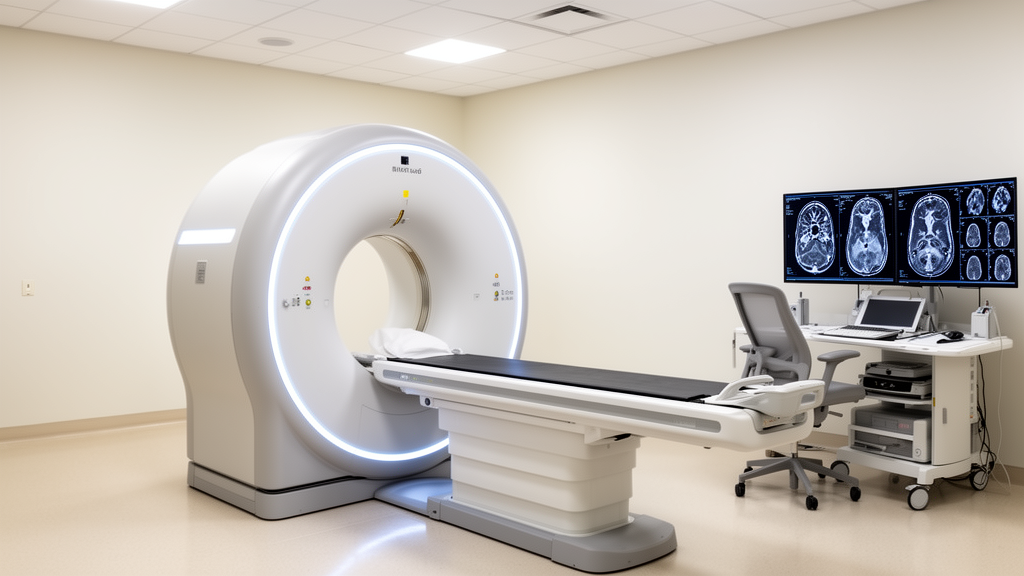
A narrative review in Frontiers in Medicine indicates that spectral computed tomography material decomposition can non-invasively quantify metabolic and elemental alterations in liver cirrhosis, expanding radiological assessment beyond structural morphology into tissue composition. Chronic liver disease represents a substantial global burden causing an average annual mortality exceeding one million individuals worldwide. Cirrhosis is not merely a localized lesion, but a systemic disorder involving disturbances across hemodynamics, immune function, and cellular metabolism. In routine clinical workflows, clinicians evaluate structural remodeling and portal hypertension complications, yet concurrent metabolic and elemental disruptions remain underestimated despite their influence on disease progression and patient survival. Conventional single-energy computed tomography is fundamentally constrained by depicting only morphological changes and general tissue attenuation. In contrast, spectral computed tomography exploits energy-dependent differences in X-ray attenuation across tissue elements, enabling simultaneous, single-scan non-invasive characterization of critical hepatic components including iron, fat, calcium, and iodine.
The physical framework of spectral computed tomography relies on acquiring attenuation datasets at two or more effective photon energy spectra to extract substance-specific information. Within diagnostic energy ranges, the linear attenuation coefficient of any tissue constituent is governed by its elemental atomic number, physical electron density, and photon energy through the photoelectric effect and Compton scattering. High-atomic-number elements, including iron, calcium, and iodine, display marked photoelectric attenuation that scales steeply with atomic number and falls rapidly as photon energies increase, whereas Compton scattering depends primarily on electron density with comparatively weak energy dependence. Spectral scanners utilize these distinct attenuation profiles to execute base material pair decompositions, generating material density images such as iron-water, calcium-water, and fat-soft tissue pairs alongside virtual single-energy reconstructions. Clinicians place regions of interest over target liver parenchyma to calculate material concentrations in milligrams per milliliter or percentage fractions, though quantification remains an estimated measurement conditioned on calibration, decomposition models, and spectral separation.
Disrupted iron homeostasis represents a major metabolic complication in progressive cirrhosis, accelerating organ failure through destructive biochemical cascades. The liver normally functions as the primary reservoir for iron storage and maintains systemic iron balance by synthesizing and secreting hepcidin, which strictly controls intestinal absorption and reticuloendothelial release. In cirrhotic parenchyma, hepatocyte necrosis and functional deterioration markedly reduce hepcidin production, prompting uncontrolled intestinal iron uptake and abnormal iron release from Kupffer cells. This unregulated intrahepatic iron accumulation drives the Fenton reaction, generating reactive oxygen species that inflict oxidative damage on hepatocyte mitochondria and persistently activate hepatic stellate cells. This harmful mechanism establishes a self-perpetuating cycle of iron overload, oxidative stress, and accelerated fibrogenesis that substantially elevates the risk of hepatocellular carcinoma. While percutaneous liver biopsy remains the traditional reference standard for quantifying hepatic iron concentration, its invasive nature, vulnerability to sampling errors, and low patient acceptance restrict its practical utility for longitudinal clinical monitoring.
To establish non-invasive surveillance, researchers developed virtual iron content imaging through spectral computed tomography utilizing iron-water decomposition algorithms to quantify hepatic iron deposition directly. Although the K-shell binding energy of iron stands at 7.1 kiloelectronvolts—below the diagnostic spectrum of conventional scanners—iron’s medium-to-high atomic number creates attenuation shifts between dual-energy acquisitions that substantially exceed ordinary soft tissues. A retrospective investigation by Werner and colleagues evaluating thoracic dual-spectral scans from 147 hematological patients confirmed that hepatic virtual iron content measurements correlated significantly with serum ferritin concentrations (r = 0.623, P < 0.001) and cumulative transfused iron volume (r = 0.558, P < 0.001). Furthermore, virtual iron content avoids the signal saturation ceilings observed during magnetic resonance R2* relaxometry in severe iron overload. Nevertheless, acquisition timing introduces technical vulnerability; overlapping attenuation between iodine and iron causes contrast-enhanced acquisitions to misattribute iodine signals to iron, producing a phase-dependent overestimation that makes unenhanced scanning the preferred clinical protocol.
Although elevated hepatic iron deposition demonstrates a strong association with advancing fibrosis, researchers emphasize that virtual iron content cannot presently serve as an independent tool for histological fibrosis staging. Hepatic iron accumulation is heavily modulated by confounding variables, including underlying disease etiology, genetic metabolic traits, repeated blood transfusions, systemic inflammation, and patient nutritional status, preventing a uniform linear relationship with histological stages. Furthermore, the frequent coexistence of hepatic steatosis and iron overload in metabolic dysfunction-associated steatotic liver disease introduces physical crosstalk that impairs dual-substance decomposition algorithms. In vitro investigations by Xie and colleagues demonstrated that an iron-water model underestimates hepatic iron concentrations within a fatty background, whereas an iron-fat model systematically overestimates iron in normal hepatic parenchyma. To overcome these mutual signal distortions, advanced three-material decomposition techniques combining iron, fat, and soft tissue have been implemented, resolving multi-substance interference and providing improved quantitative precision across complex hepatic tissue environments where fat and iron simultaneously accumulate.
Disorders of calcium metabolism and secondary hepatic bone disease represent another critical yet frequently neglected complication in cirrhotic populations that spectral imaging may help elucidate. Calcium homeostasis depends on coordinated interactions among intestinal absorption, renal excretion, and skeletal reserves, with the liver acting as the indispensable metabolic clearinghouse that hydroxylates vitamin D into 25-hydroxyvitamin D. In cirrhotic patients, impaired hepatocyte integrity suppresses 25-hydroxylase enzymatic activity, producing severe 25-hydroxyvitamin D deficiencies, blunted intestinal calcium uptake, and secondary hyperparathyroidism that mobilizes skeletal calcium and accelerates bone loss. Epidemiological reviews by Korkmaz and colleagues indicate that osteoporosis affects between 12% and 55% of individuals with advanced chronic liver disease, producing a two- to four-fold higher fracture risk and causing vertebral fractures in 20% to 30% of cirrhotic patients. A study by Jamil and colleagues demonstrated that 88% of chronic liver disease patients exhibited vitamin D insufficiency or deficiency, with serum levels inversely correlating with Child-Pugh class.
Despite the recognized clinical severity of systemic bone mineral loss, the quantitative evaluation of intrahepatic calcium accumulation via spectral computed tomography remains confined to theoretical models and preliminary laboratory experiments. Technical validation of calcium-water material decomposition conducted by Miller and colleagues using standardized physical phantoms demonstrated that accurate calcium measurement requires disabling iodine-specific beam hardening corrections and applying size-specific dual-energy ratios, while computed tomography dose index and iterative reconstruction intensity do not significantly distort measurements. Virtual calcium images theoretically allow direct measurement of parenchymal calcium density, yet clinical investigations confirming this capability in human cirrhotic cohorts are entirely lacking. Inherent physical limitations present substantial obstacles: physiological baseline calcium concentrations in hepatic parenchyma are extremely low, and calcium possesses a low K-shell binding energy of 4.0 kiloelectronvolts, generating subtle attenuation variations that yield detection sensitivity far inferior to iron quantification, while diverse systemic factors like diet and renal function complicate clinical calibration.
In contrast to calcium, non-invasive quantification of hepatic steatosis via spectral computed tomography is substantially more advanced and exhibits strong technical alignment with magnetic resonance imaging proton density fat fraction. Fat quantification exploits the pronounced attenuation disparity between adipose tissue and hepatic soft tissue across varied energy spectra, utilizing multimaterial decomposition algorithms to calculate hepatic fat volume fractions. A comprehensive 2024 meta-analysis by Haghshomar and colleagues analyzing 42 studies comprising 14,186 participants identified pooled sensitivities of 72% for non-contrast computed tomography and 84% for dual-energy scans in detecting any steatosis, with specificities of 88% and 93% respectively; however, when dual-energy scans were compared directly against liver biopsy or magnetic resonance imaging proton density fat fraction, pooled specificity was 88%, matching non-contrast scanning and demonstrating that dual-energy computed tomography is not conclusively superior to standard non-contrast imaging. Furthermore, comparative studies by Hyodo and colleagues revealed that multimaterial decomposition measurements correlate closely with magnetic resonance spectroscopy, with equilibrium-phase contrast scans displaying close agreement with unenhanced acquisitions.
While iron and lipid imaging systems have established measurable clinical utility, direct in vivo quantification of vital trace elements such as zinc and copper remains obstructed by insurmountable physical boundaries under routine spectral computed tomography. Systemic zinc deficiency occurs extensively across cirrhotic cohorts due to impaired intestinal absorption driven by portal hypertension, hypoalbuminemia, and parenchymal damage, thereby worsening hepatic encephalopathy, immune suppression, and delayed wound repair. Conversely, impaired biliary clearance causes pathological copper retention within cirrhotic tissue, catalyzing oxidative parenchymal injury through harmful Fenton-like chemical cascades. Despite the clinical necessity of monitoring these metabolic perturbations, conventional spectral computed tomography cannot directly differentiate or quantify trace zinc and copper concentrations. This technical barrier exists because the K-edge absorption energies of zinc at 9.66 kiloelectronvolts and copper at 8.98 kiloelectronvolts reside far below the 50 to 80 kiloelectronvolt effective operating range of diagnostic scanners, while minute physiological concentrations prevent routine clinical decomposition algorithms from extracting detectable elemental signals.
Overcoming these diagnostic bottlenecks will depend heavily on emerging technical architectures that integrate photon-counting computed tomography with deep learning reconstruction models. Unlike conventional energy-integrating detectors, photon-counting computed tomography uses direct-conversion semiconductor sensors to measure individual photons and categorize them into multiple energy bins, thereby lowering energy thresholds toward zinc and copper K-edges while reducing image noise and improving spatial resolution. Simultaneously, deep learning algorithms can decipher complex non-linear attenuation relationships across multi-energy acquisitions, bypassing the mathematical simplifications inherent in traditional basis-pair models. Before these advanced quantitative tools can transition into routine clinical environments, however, significant operational hurdles must be resolved, including harmonizing disparate manufacturer platforms, standardizing region-of-interest placements, and establishing protocol-specific calibration standards. Until rigorous multicenter clinical trials confirm their diagnostic accuracy across diverse patient cohorts, researchers suggest constructing correlation models that link robustly measured iron, fat, and calcium indices with underlying trace element dynamics to better characterize the multidimensional metabolic disruptions of cirrhosis.
Subject of Research: Spectral computed tomography material decomposition for metabolic and elemental quantification in liver cirrhosis
Article Title: Spectral CT material decomposition for non-invasive quantitative assessment of metabolic and elemental abnormalities in liver cirrhosis: a narrative review
Article References: Zhang, R., Li, Z., Lin, T., Li, Y., Su, C., Song, H., & Du, T. (2026). Spectral CT material decomposition for non-invasive quantitative assessment of metabolic and elemental abnormalities in liver cirrhosis: a narrative review. Frontiers in Medicine, 13, 1988368. https://doi.org/10.3389/fmed.2026.1988368
Image Credits: AI Generated
DOI: 10.3389/fmed.2026.1988368
Keywords: spectral CT, material decomposition, liver cirrhosis, virtual iron content, hepatic steatosis, multimaterial decomposition, MRI-PDFF, photon-counting CT, hepatic bone disease, trace elements, zinc deficiency, copper accumulation, dual-energy CT, Fenton reaction, hepcidin, deep learning CT, calcium-water decomposition, iron overload
News Source: Ophelia Keating. (November 11, 2026). Spectral CT Quantifies Hepatic Iron and Fat but Misses Trace Elements. Scienmag.