Anemia remains one of the most pervasive global health challenges, impacting nearly two billion individuals worldwide. Particularly affected are school-age children in low- and middle-income countries where healthcare infrastructure often falls short. The condition, characterized by deficient hemoglobin levels in the blood, can profoundly disrupt physical growth, cognitive development, and academic performance if not diagnosed and treated early. Current gold-standard detection techniques necessitate invasive blood draws and laboratory analysis—procedures impractical in many resource-limited settings. However, a groundbreaking study spearheaded by researchers across Purdue University, Rwanda Biomedical Center, and University of Rwanda proposes an innovative, noninvasive diagnostic method that harnesses the power of machine learning combined with radiomic image analysis, offering a new frontier for anemia detection.
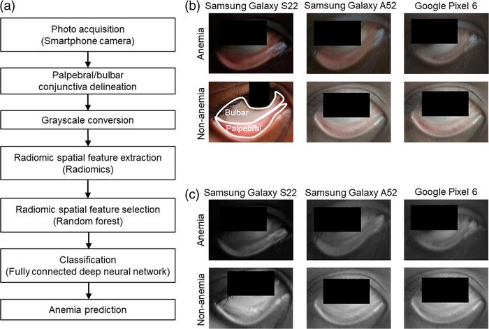
The novel approach capitalizes on the detailed examination of grayscale photographs of the conjunctiva, the delicate mucous membrane lining the inner eyelid and visible sclera, captured using commonplace smartphone cameras. Radiomics, a cutting-edge analytical technique, extracts quantitative mathematical descriptors related to spatial arrangement and texture from medical images, unveiling subtle patterns imperceptible to the human eye. Over 12,000 eye images were collected from 565 children aged between five and fifteen years, forming the largest dataset of its kind in this context. This extensive image repository underpins a machine learning model trained to identify radiomic signatures associated with anemia status, sidestepping the need for color detection algorithms traditionally used in similar efforts.
Shaun Hong, the first author and a PhD candidate at Purdue, emphasizes the significance of eschewing color in this model. “Our method bypasses dependence on color data and instead focuses on the microstructural variations in conjunctival blood vessels captured in black-and-white imagery,” he explains. This pivotal decision mitigates confounding factors such as inconsistent lighting environments and the spectral variability arising from diverse smartphone camera sensors. The strategy ensures robustness and adaptability, expanding applicability across disparate geographic regions and device types.
The study’s results unveil a compelling correlation between specific radiomic features extracted from the monochromatic conjunctiva photos and the presence of anemia. Textural heterogeneity, spatial irregularities in vascular patterns, and subtle morphological markers embedded in the grayscale imagery serve as robust predictors of hemoglobin deficiency. This finding underscores the potential of leveraging minimalistic yet rich image data through sophisticated computational algorithms for impactful clinical screening.
Beyond theoretical validation, this research carries profound practical implications. Conventional anemia diagnostics remain constrained by the need for phlebotomy and laboratory infrastructure, often unavailable or unreliable in remote communities. A smartphone-based, image-driven diagnostic tool configured with embedded radiomic and machine learning models could facilitate immediate, on-site anemia risk assessment. Such innovation portends transformative shifts in public health screening protocols, especially in resource-constrained environments where traditional laboratory tools are infeasible.
Professor Young L. Kim of Purdue University, a corresponding author on the study, notes the envisioned clinical utility: “Our technology is not intended to supplant laboratory diagnostics but to serve as an adjunct prioritization mechanism. Enabling frontline health workers to swiftly screen children remotely helps direct scarce resources to patients who most urgently require confirmatory testing and intervention.” This triage capacity is invaluable in maximizing healthcare efficiency and improving early outcomes through targeted management.
Radiomics itself represents an intersection of imaging science, data analytics, and clinical insight. By translating complex textural and spatial image properties into quantitative data points, radiomics empowers machine learning algorithms to recognize disease-specific phenotypes invisible to naked observation. In this context, careful pre-processing of images ensures standardization, and feature selection algorithms optimize the model’s predictive accuracy by isolating the most relevant radiomic markers linked to anemia-induced conjunctival changes.
The utilization of standard smartphones in this research also heralds democratization of diagnostic technology. Most mobile devices today are equipped with sufficiently capable cameras and processing power to capture and analyze medical images, facilitated by readily deployable software applications. This scalability could enable thousands of frontline clinics, school health programs, and community health initiatives worldwide to implement digital anemia screening protocols at negligible incremental cost, bypassing logistical bottlenecks inherent to traditional testing.
Furthermore, this technique’s noninvasiveness enhances patient compliance, particularly among children, who may otherwise find blood sampling intimidating or inaccessible. Reporting turnaround time shrinks dramatically, allowing near real-time decision-making that can immediately influence clinical pathways. Such expedient care delivery is critical in mitigating anemia’s chronic adverse effects on child development and educational attainment.
The research team’s collaborative international effort underscores the integration of diverse expertise spanning biomedical engineering, epidemiology, and computational science. Their work, published in the open-access journal Biophotonics Discovery, presents a compelling proof-of-concept supported by rigorous validation metrics and promising pathways for future refinement. As the model matures, incorporation of additional biometric inputs and cross-population validation will likely enhance generalizability and robustness.
In the broader landscape, this study exemplifies how emerging digital health technologies, fused with artificial intelligence and image analysis, can surmount longstanding clinical barriers in low-resource settings. The strategic use of radiomics to decode monochromatic image data places this work at the forefront of innovative disease detection paradigms, pointing towards a future where smartphone-enabled diagnostics become an indispensable tool in global health.
Ultimately, by unlocking the potential of simple eye photographs analyzed through sophisticated radiomic and machine learning frameworks, this research paves the way for scalable, affordable, and noninvasive anemia screening solutions. Such advancements contribute critically to reducing the global anemia burden, supporting healthier development trajectories for vulnerable children worldwide, and advancing equitable access to quality healthcare diagnostics.
Subject of Research: Anemia detection using radiomic analysis of monochromatic conjunctiva photographs in school-age children
Article Title: Radiomic identification of anemia features in monochromatic conjunctiva photographs in school-age children
News Publication Date: 15-Apr-2025
Web References: https://www.spiedigitallibrary.org/journals/biophotonics-discovery/volume-2/issue-02/022303/Radiomic-identification-of-anemia-features-in-monochromatic-conjunctiva-photographs-in/10.1117/1.BIOS.2.2.022303.full
References: S. G. Hong et al., “Radiomic identification of anemia features in monochromatic conjunctiva photographs in school-age children,” Biophotonics Discovery 2(2), 022303 (2025), doi: 10.1117/1.BIOS.2.2.022303
Image Credits: S. S. Hong et al., 10.1117/1.BIOS.2.2.022303
Keywords: Anemia, Smartphones, Machine learning, Radiomics, Conjunctiva, Hemoglobin, Image analysis, Medical technology, Blood diseases, Computational biology
Tags: anemia detection in childrenconjunctiva imaging technologyglobal health challengesinnovative health solutionslow-income country healthcaremachine learning in healthcarenoninvasive diagnostic methodspediatric anemia diagnosisquantitative medical image analysisradiomic image analysisresource-limited healthcare settingssmartphone eye imaging


