In a retrospective comparative-effectiveness evaluation, extracorporeal shock wave therapy demonstrated greater short-term improvements in pain and shoulder function than conventional exercise-based physical therapy among patients presenting with noncalcific rotator cuff tendinopathy or partial-thickness tears. The investigation, conducted at the Wucheng District First People’s Hospital of Jinhua City and published in Frontiers in Medicine, tracked forty-four adult patients across six weeks of clinical care. Beyond establishing functional and symptomatic comparisons across standardized clinical metrics, the clinical investigators evaluated quantitative baseline magnetic resonance imaging via T2 relaxation mapping to determine whether native tendon microstructure and tissue heterogeneity could correlate with short-term clinical outcomes following radial shock wave intervention.
Rotator cuff disorders represent a frequent etiology of shoulder pain and disability, encompassing a clinical spectrum that spans tendinopathy without definite fiber discontinuity to partial-thickness and full-thickness tendon tears. While nonoperative management typically relies on conservative protocols such as analgesics, therapeutic ultrasound, and structured physical rehabilitation, radial extracorporeal shock wave therapy has emerged as an alternative mechanical intervention. Standard diagnostic workups depend heavily on conventional magnetic resonance imaging, including T1-weighted and fat-suppressed proton density-weighted sequences, to visualize macrostructural tendon morphology, tear dimensions, and tendon retraction. However, standard qualitative MRI offers limited direct quantification of soft-tissue biochemistry, moisture content, collagen architecture, or internal material properties, prompting interest in quantitative magnetic resonance techniques such as T2 mapping.
Quantitative T2 mapping measures tissue T2 relaxation times in milliseconds, which reflect biochemical compositions including water content and collagen fiber organization within musculoskeletal tissues. In healthy and degenerated supraspinatus tendons, altered collagen integrity and elevated tissue water concentration lead to shifts in measurable relaxation times, with pathologic tear zones often exhibiting higher regional T2 values than intact or asymptomatic tendon areas. In this investigation, the clinical team sought to resolve two distinct research questions: first, whether patients undergoing radial shock wave therapy achieved superior short-term pain relief and functional restoration compared with conventional physical rehabilitation; and second, whether baseline tendon T2 values or spatial heterogeneity could serve as exploratory imaging correlates of clinical improvement among individuals receiving shock wave therapy.
The study cohort was drawn from forty-nine consecutive patients screened between January and December 2023 who presented with persistent shoulder symptoms confirmed by magnetic resonance imaging. Individuals with calcific tendinopathy, full-thickness tendon disruptions, prior shoulder operations, fractures, severe joint deformities, concurrent neurological conditions affecting upper-limb movement, or systemic contraindications such as bleeding coagulopathies were excluded, leaving forty-four eligible participants. Allocation to treatment was determined during routine clinical practice based on physician evaluation, imaging presentation, and patient choice, yielding two equal cohorts of twenty-two individuals. One cohort underwent radial extracorporeal shock wave therapy, while the comparative control group received conventional physical therapy consisting of ultrasound and progressive exercise.
Imaging protocol standardization involved acquiring high-field 3.0-Tesla shoulder MRI with a dedicated joint coil before initiating any clinical intervention. Quantitative T2 maps were constructed in the oblique coronal plane along the long axis of the supraspinatus tendon using a multiecho spin-echo sequence covering repetition times of 2000 milliseconds and echo times between 10 and 80 milliseconds. To extract objective quantitative metrics, two independent radiologists, blinded to treatment allocations and clinical trajectories, positioned circular regions of interest centered directly on the most conspicuous abnormal tendon tissue identified on matched proton density-weighted views. From these defined regions, the mean T2 value was calculated to represent overall tissue relaxation status, while the standard deviation of T2 values was computed to capture intraregional signal heterogeneity, achieving interobserver and intraobserver intraclass correlation coefficients ranging from 0.71 to 0.94.
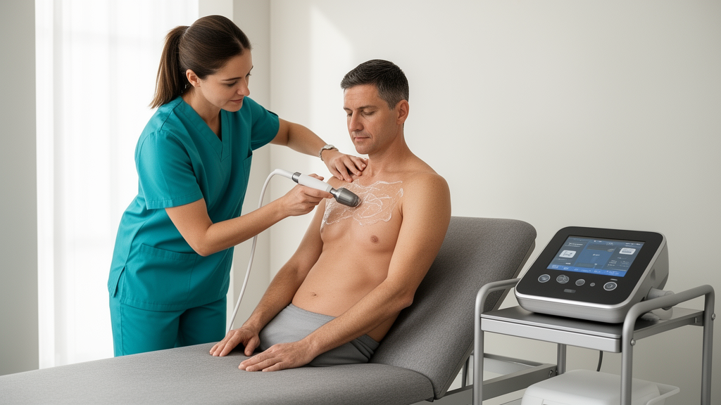
Patients assigned to the shock wave intervention received radial extracorporeal shock wave therapy using a Storz Medical MASTERPULS MP100 device applied to the MRI-identified lesion and the corresponding site of maximum clinical tenderness. Sessions were administered once weekly for three to five total visits, delivering between 1,561 and 3,202 impulses per session at native operating settings individualized to patient tolerance between 2 and 4 bar and 5 to 15 Hz. An exploratory treatment-intensity index was documented within a range of 0.19 to 0.32. Conversely, control participants completed therapeutic ultrasound delivered at 1 MHz and 1.0 to 1.2 W/cm² for five to eight minutes per session, accompanied by supervised exercise therapy conducted three to five times weekly over the six-week timeline, featuring active-assisted mobility, scapular stabilization, and rotator cuff strengthening.
Patient status was evaluated at baseline and at the conclusion of the six-week period using three established clinical instruments. Primary outcome evaluation centered on pain severity tracked via a 100-millimeter visual analogue scale and overall shoulder functional integrity assessed through the 100-point Constant-Murley Score, with the University of California, Los Angeles shoulder score monitored as a supplementary composite index. In parallel, the investigators established an operational response threshold requiring both a reduction in visual analogue scale pain of at least 50 percent and an increase in Constant-Murley functional score of at least 20 percent to classify an individual as a clinical responder.
At baseline, demographic and clinical parameters were statistically balanced between the two study arms, with mean ages of 45.1 years in the shock wave arm and 49.6 years in the physical therapy arm, comparable symptom durations of approximately 6.5 months, and similar baseline pain, functional scores, and T2 values. Following six weeks of intervention, the shock wave cohort exhibited significantly lower post-treatment pain on the visual analogue scale compared with controls, registering 42.14 points versus 59.00 points. The relative pain reduction was substantially higher in the shock wave group at 39.91 percent compared with 11.66 percent in the physical therapy cohort, and supplementary UCLA score improvements favored shock wave delivery at 5.05 points versus 1.32 points. While raw post-treatment Constant-Murley scores did not achieve statistically significant divergence in unadjusted testing, baseline-adjusted linear regression analyses demonstrated significant advantages for shock wave therapy across visual analogue scale pain, Constant-Murley function, and UCLA scores.
Exploratory evaluations within the twenty-two patients treated with shock wave therapy identified distinct quantitative imaging patterns between treatment responders and non-responders. Five patients met the operational clinical response criteria, whereas seventeen did not. Baseline measurements revealed that responders began treatment with significantly higher mean T2 values of 56.20 milliseconds compared with 34.82 milliseconds in non-responders, along with markedly elevated T2 standard deviations of 10.00 milliseconds versus 2.88 milliseconds, indicating substantial baseline tissue heterogeneity. However, responders also presented with significantly lower initial Constant-Murley scores of 26.20 points compared with 42.65 points in non-responders, demonstrating that subjects experiencing the largest percentage gains possessed greater initial functional impairment.
Correlation analyses among shock wave recipients demonstrated outcome-specific relationships between baseline imaging variables, administered technical dosage, and subsequent recovery. Baseline T2 standard deviation correlated with percentage pain reduction, whereas baseline mean T2 correlated with percentage functional improvement on the Constant-Murley scale. In baseline-adjusted sensitivity models, T2 standard deviation remained significantly associated with six-week visual analogue scale pain after controlling for initial pain scores; however, this association was attenuated when adjusting for the recorded treatment-intensity index. The treatment-intensity index itself correlated with pain reduction as well as baseline mean T2 and T2 standard deviation, indicating that administered acoustic energy and pre-existing tendon composition were not clinically independent.
The authors highlighted several critical methodological limitations inherent to the study design that constrain causal interpretations. Because treatment allocation occurred within routine clinical workflows without randomization, selection bias and confounding by indication cannot be excluded. Furthermore, the overall sample size of forty-four patients produced small subgroup numbers, yielding wide confidence intervals around binary responder analyses. Physical therapy served as an active clinical comparator rather than a sham-shockwave procedure, meaning non-specific contextual factors and unequal clinical contact times could not be isolated. Additionally, the clinical examiners were aware of treatment assignments, the operational composite responder definition lacked external validation, and the follow-up window was restricted to six weeks, precluding long-term assessment of structural tissue durability.
Subject of Research: Extracorporeal shock wave therapy and quantitative MRI T2 mapping in rotator cuff lesions
Article Title: Short-term clinical outcomes of extracorporeal shock wave therapy and exploratory associations with baseline MRI T2 mapping in rotator cuff lesions: a retrospective study
Article References: Zheng, P., & Huang, W. (2026). Short-term clinical outcomes of extracorporeal shock wave therapy and exploratory associations with baseline MRI T2 mapping in rotator cuff lesions: a retrospective study. Frontiers in Medicine, 13, Article 1939434. https://doi.org/10.3389/fmed.2026.1939434
Image Credits: AI Generated
DOI: 10.3389/fmed.2026.1939434
Keywords: extracorporeal shock wave therapy, rotator cuff tendinopathy, partial-thickness tear, MRI T2 mapping, visual analogue scale, Constant-Murley Score, UCLA shoulder score, musculoskeletal imaging, physical therapy, shoulder rehabilitation, tendon microstructure, comparative effectiveness, Storz Medical MASTERPULS MP100, therapeutic ultrasound, exercise therapy, collagen organization
News Source: Ophelia Keating. (November 21, 2026). Shock Wave Therapy Eases Shoulder Tendon Pain in Comparative Clinical Study. Scienmag.