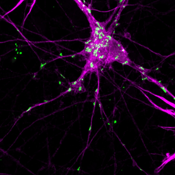
A novel system to control protein aggregation in a model of Parkinson’s disease may answer longstanding questions about how the disease begins and spreads, according to a new study published March 9th in the open-access journal PLOS Biology by Abid Oueslati of Laval University, Quebec, Canada, and colleagues. Initial results suggest that aggregation of the protein alpha-synuclein plays a critical role in disrupting neuronal homeostasis and triggering neurodegeneration.

Credit: Maxime Teixeira (CC-BY 4.0, https://creativecommons.org/licenses/by/4.0/)
A novel system to control protein aggregation in a model of Parkinson’s disease may answer longstanding questions about how the disease begins and spreads, according to a new study published March 9th in the open-access journal PLOS Biology by Abid Oueslati of Laval University, Quebec, Canada, and colleagues. Initial results suggest that aggregation of the protein alpha-synuclein plays a critical role in disrupting neuronal homeostasis and triggering neurodegeneration.
Parkinson’s disease is a neurodegenerative disorder, marked clinically by tremor, stiffness, and slowed movements, as well as a host of nonmotor symptoms. Within affected neurons, molecules of a protein called alpha-synuclein can be seen to clump together, forming characteristic aggregates called Lewy bodies. But it has been hard to answer whether alpha-synuclein aggregation contributes to disease development or progression, and when it may act in the toxic disease cascade, or whether instead the aggregates are innocent bystanders to some other malevolent process, or are even protective. These elements have been difficult to determine, in part because aggregation in cellular and animal models has not been controllable in either time or space.
To address that problem, the authors turned to optobiology, a technique in which a protein of interest is fused to another protein that changes its conformation in response to light, allowing the behavior of the target protein to be manipulated selectively and reversibly. Here, the authors fused alpha-synuclein to a protein known as cryptochrome protein 2, from a mustard plant. They found that when light of the correct wavelength fell on the mustard protein, its conformational change triggered aggregation of its alpha-synuclein partner.
The aggregates that formed were reminiscent of Lewy bodies in multiple important ways, including that they included several other key proteins besides alpha-synuclein found in Lewy bodies in people with Parkinson’s disease, and that the alpha-synuclein in the aggregates adopted the characteristic beta-sheet conformation seen in many diseases of misfolded proteins. The aggregates induced dislocation of multiple cellular organelles, as Lewy bodies have been recently reported to do as well. They also induced misfolding in alpha-synuclein molecules not attached to the cryptochrome protein, mimicking the prion-like spread of aggregation seen with alpha-synuclein in the diseased brain and animal models.
Finally, the authors delivered the genes for the alpha-synuclein-cryptochrome fusion protein to mice, directly into the substantia nigra, the structure in the brain that is most prominently affected by Parkinson’s disease, and surgically placed an optic fiber to deliver light to the targeted cells. Light treatment led to formation of alpha-synuclein aggregates, neurodegeneration, disruption of calcium activity in downstream neuronal targets, and Parkinson-like motor deficits.
“Our results demonstrate the potential of this optobiological system to reliably and controllably induce formation of Lewy body-like aggregations in model systems, in order to better understand the dynamics and timing of Lewy body formation and spread, and their contribution to the pathogenesis of Parkinson’s disease,” Oueslati said.
Oueslati adds, “How do alpha-synuclein aggregates contribute to neuronal damage in Parkinson’s disease? To help address this question, we developed a new optogenetic-based experimental model allowing for the induction and real-time monitoring of alpha-synuclein clustering in vivo.”
#####
In your coverage, please use this URL to provide access to the freely available paper in PLOS Biology: http://journals.plos.org/plosbiology/article?id=10.1371/journal.pbio.3001578
Citation: Bérard M, Sheta R, Malvaut S, Rodriguez-Aller R, Teixeira M, Idi W, et al. (2022) A light-inducible protein clustering system for in vivo analysis of α-synuclein aggregation in Parkinson disease. PLoS Biol 20(3): e3001578. https://doi.org/10.1371/journal.pbio.3001578
Author Countries: Canada, United States, Republic of Korea
Funding: This work was supported by Parkinson Society Canada, Fondation du CHU de Québec and the Canadian Institutes of Health Research (CIHR) grants to AO. AO was supported by Junior1 and Junior 2 salary Awards from the Fonds de Recherche du Québec – Santé (FRQS) and la Société Parkinson du Québec. In vivo Ca2+ imaging experiments were supported by the Canadian Institutes of Health Research (CIHR) grant to AS. MB was supported by scholarships from the Fondation du CHU de Québec, Faculty of Medicine of Université Laval (Bourse de recrutement du doctorat Pierre J. Durant), and FRQS. FC is a recipient of a Researcher Chair from the Fonds de Recherche du Québec en Santé (FRQS) and received funding from the Canadian Institutes of Health Research (CIHR). MA is supported by post-doctoral fellowships from both CIHR and FRQS. E.A.F. is supported by a Foundation grant from CIHR (FDN-154301) and a Canada Research Chair (Tier 1) in Parkinson’s disease. MKSP was supported by a Frederick Banting and Charles Best Canada Graduate Scholarship-Doctoral Award and a Doctoral’s training scholarship from FRQS. MET is a Tier 2 Canada Research Chair in Neurobiology of Aging and Cognition. TMD is supported with funds from the McGill Healthy Brains for Healthy lives and a project grant from CIHR (PJT–169095). The funders had no role in study design, data collection and analysis, decision to publish, or preparation of the manuscript.
Journal
PLoS Biology
DOI
10.1371/journal.pbio.3001578
Method of Research
Experimental study
Subject of Research
Cells
COI Statement
Competing interests: The authors have declared that no competing interests exist.


