


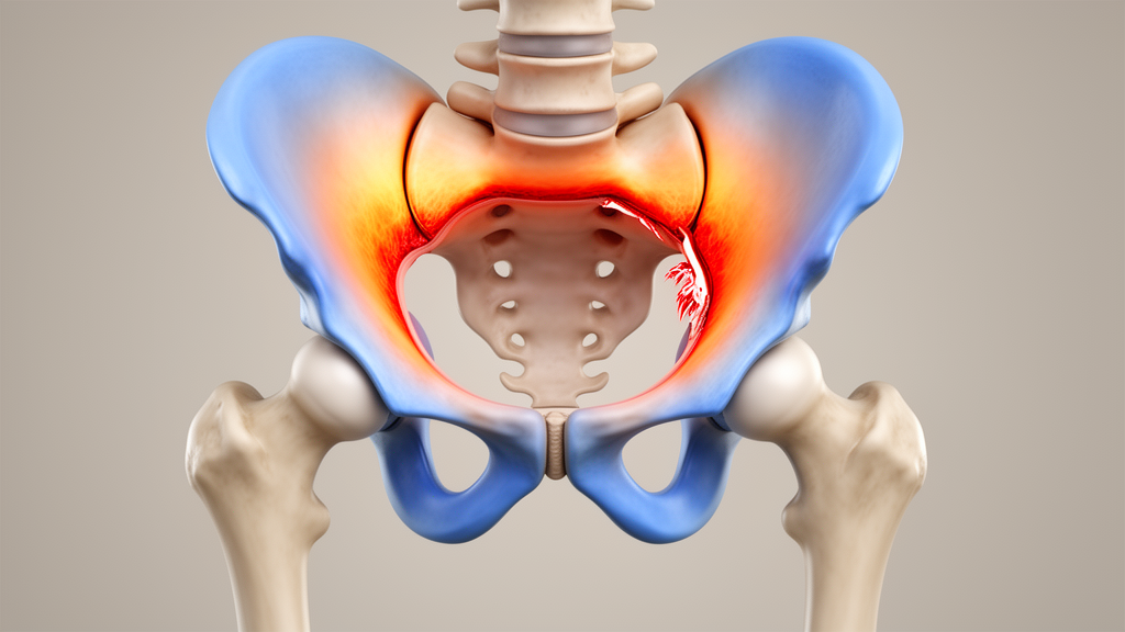
Women with developmental dysplasia of the hip who carry complicated labral injuries show a striking pattern of stress concentration in the upper acetabulum, according to a cross-sectional study published in Archives of Orthopaedic and Trauma Surgery. Using computed tomography osteoabsorptometry, a technique that maps subchondral bone density as a proxy for cumulative joint loading, researchers found that the proportion of high-density bone in the superior acetabulum rose from 64.4 percent in hips with simple labral injuries to 80.3 percent in those with complicated injuries, a difference that was highly significant. The finding links the severity and location of labral damage to a measurable redistribution of long-term mechanical load within the joint.
Developmental dysplasia of the hip is a morphological abnormality in which the acetabulum provides shallow coverage of the femoral head. This geometry concentrates loading stress at the edge of the joint and predisposes the labrum, a ring of fibrocartilage that helps seal the joint and stabilise the femoral head, to damage. Finite element studies cited by the authors indicate that in a normal hip the labrum carries only one to two percent of the total applied load, whereas in dysplastic hips this share rises to four to eleven percent. Athletes with dysplasia, including ballet dancers and long-distance runners, face an elevated risk of labral injury because repetitive hip movements multiply the mechanical stress on an already vulnerable structure.
Labral injuries are common in this population. Among symptomatic patients with dysplasia who require surgical intervention, tears or degeneration of the labrum have been reported in roughly ninety percent of cases. Injuries can occur anywhere around the joint circumference, but they cluster in the anterior region, followed by the superior region. What has remained unclear is the direction of the relationship between labral damage and joint loading: whether the injury drives an abnormal distribution of stress, or whether abnormal stress distribution is what damages the labrum in the first place. Resolving this question matters clinically, because capsular laxity in dysplastic hips means that arthroscopy can worsen instability, and the value of intra-articular labral interventions in these patients remains debated.
The study team, led by Yuki Ogawa and colleagues, analysed 54 hips from 45 female patients who underwent joint-preserving periacetabular osteotomy for dysplasia at a single centre between 2016 and 2024. The cohort was restricted to women because only four male patients were identified, an imbalance too marked to evaluate sex-related differences. Patients averaged 30.3 years of age and a body mass index of 21.7 kilograms per square metre. Before surgery, each hip underwent radial magnetic resonance imaging, in which 18 slices were acquired at ten-degree intervals perpendicular to the acetabular labrum, and plain computed tomography for bone density mapping and finite element modelling.
On the radial MRI scans, labral lesions were classified into two severity categories. Simple injuries comprised abnormal intralabral signals or simple tears confined to one site, with tears spanning several subregions in a single direction still counted as simple. Complicated injuries involved complex tears extending in multiple directions or maceration of the labrum. For each of the three acetabular regions, anterior, superior and posterior, the most severe finding among the radial slices defined that region’s representative grade. Interobserver agreement for this classification was 0.88 and intraobserver agreement 0.90, indicating strong reliability.
Computed tomography osteoabsorptometry quantified subchondral bone mineral density in Hounsfield units along the acetabular arc, with calibration phantoms scanned alongside patients to standardise values. The high-density area was defined as the region containing the top twenty percent of density values across the articular surface, and its percentage within each acetabular region served as the measure of where cumulative load had accumulated over time. Because subchondral bone remodels in response to the stresses it experiences, this density map is read as a record of long-term loading rather than a snapshot of a single moment. Reliability was high, with intraobserver and interobserver correlation coefficients of 0.92 and 0.89 respectively.
The comparison between simple and complicated superior labral injuries revealed the study’s central result. Hips with complicated superior injuries had a significantly higher superior high-density area percentage than those with simple injuries, 80.3 percent versus 64.4 percent, while the anterior high-density percentage was significantly lower in the complicated group. In a linear mixed-effects regression that accounted for bilateral hips within patients and included age, center-edge angle, acetabular roof obliquity and the presence of complicated injury in each region as predictors, complicated superior labral injury emerged as a positive influencing factor for superior high-density percentage. No significant correlations were found for the anterior or posterior high-density percentages, and complicated anterior or posterior injuries did not significantly influence density in their own regions.
Finite element analysis told a different story. Three-dimensional models of each patient’s pelvis and femur, built from CT data with cartilage modelled as a uniform 1.8-millimetre layer, were loaded under a simulated single-leg stance with a standardised 1158-newton force applied at the femoral head centre. Nonlinear contact analysis found no significant difference between the simple and complicated superior injury groups in maximum contact pressure or contact area, with pressures of 5.3 versus 6.2 newtons per square millimetre and a p value of 0.538. The authors interpret this null result as evidence that bony morphology alone cannot explain the density patterns observed, since the models deliberately excluded the labrum itself, whose complex anisotropic fibrocartilaginous properties make reliable modelling infeasible with current techniques.
The discrepancy between the two methods is central to the paper’s argument. Finite element analysis captures instantaneous stresses under a single static posture, whereas subchondral bone density reflects the cumulative history of repetitive loading during daily activities. The authors therefore suggest that labral failure contributes to abnormal load distribution in ways that bone geometry does not capture, a possibility supported by cadaveric biomechanical studies showing that labral disruption increases intra-articular stress. They also note that the labrum is richly innervated and that labral injury has been reported to alter neuromuscular control and movement patterns, which could further modify how loads are distributed during dynamic activity. Whether the association reflects a cause or a consequence of abnormal loading cannot be determined from a cross-sectional design.
Several limitations frame these conclusions. Stress distribution was not measured directly but inferred from bone density, which may not fully reflect absolute stress values, although patients with acetabular bone cysts that could distort measurements were excluded. The cohort consisted of symptomatic surgical candidates, so the frequency and severity of labral injuries may be overestimated relative to the broader dysplastic population, and intact labra were so rare that comparisons with uninjured hips were impractical. A uniform body weight was assumed in the finite element models to remove scaling effects, and some subgroup comparisons involved small numbers, so the absence of significant finite element differences may reflect Type II error. Finally, only female patients were studied, and the associations may not extend to men. Within those bounds, the study demonstrates that complicated superior labral injuries in women with hip dysplasia are associated with localised stress accumulation in the superior acetabulum, a pattern not explained by bone shape alone, while anterior injuries, though the most frequent, appear driven more by the joint’s inherent structure than by progressive stress accumulation.
Subject of Research: The association between labral injury severity and subchondral bone density patterns indicating stress distribution in dysplastic hips
Article Title: Complicated labral injuries in female patients with developmental dysplasia of the hip are associated with localized stress concentration in the superior acetabulum
Article References: Ogawa, Y., Shimizu, T., Ohashi, Y., Sugawara, Y., Takahashi, D., & Iwasaki, N. (2026). Complicated labral injuries in female patients with developmental dysplasia of the hip are associated with localized stress concentration in the superior acetabulum. Archives of Orthopaedic and Trauma Surgery, 146(1), Article 377. https://doi.org/10.1007/s00402-026-06452-8
Image Credits: AI Generated
DOI: 10.1007/s00402-026-06452-8
Keywords: developmental dysplasia of the hip, acetabular labrum, labral injury, subchondral bone density, computed tomography osteoabsorptometry, finite element analysis, joint stress distribution, radial MRI, periacetabular osteotomy, hip biomechanics, Complicated, labral
News Source: Ophelia Keating. (October 28, 2026). Severe labral tears in dysplastic hips concentrate stress in the upper acetabulum. Scienmag.



