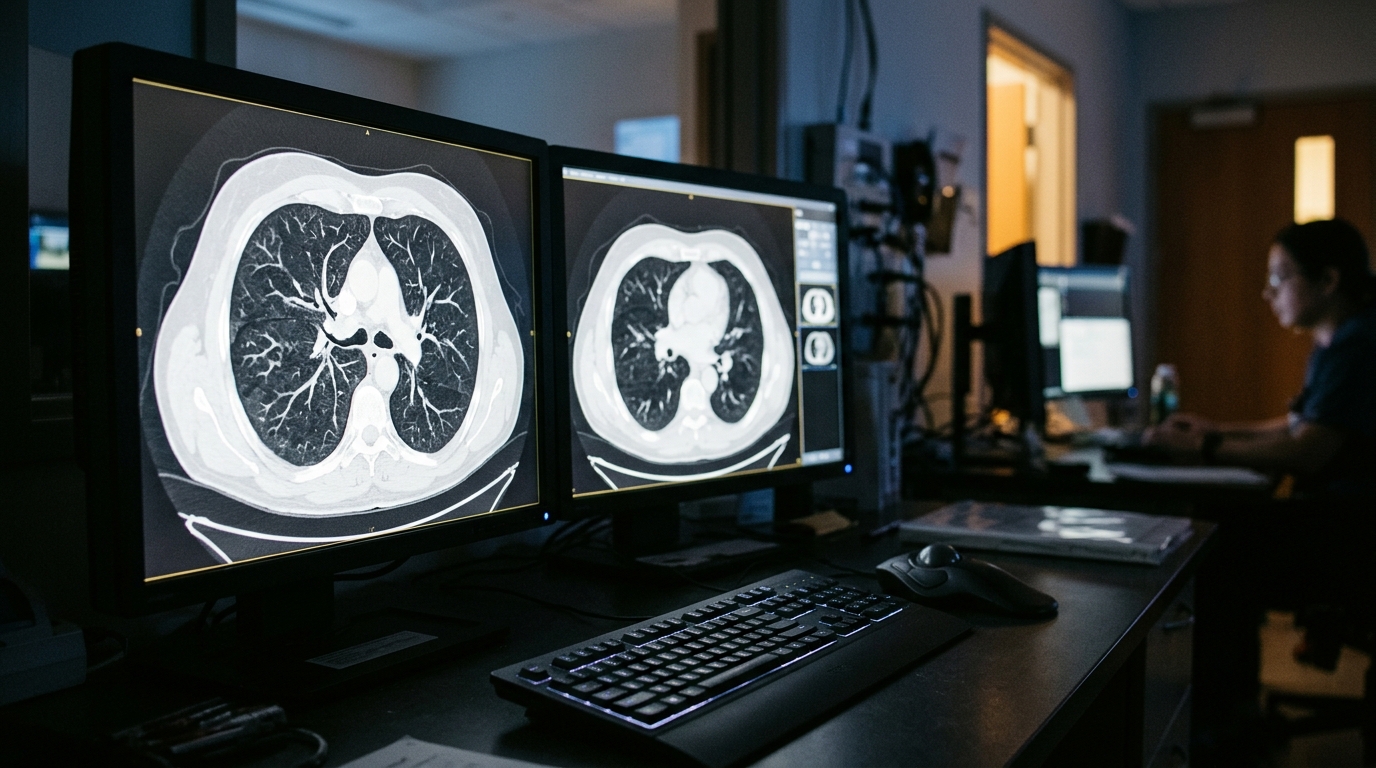
Sequential computed tomography imaging has tracked the longitudinal development of primary pulmonary mesenchymal chondrosarcoma originating from the wall of a pulmonary cyst, documenting tumor changes across a 14-month timeframe in a published case report.
Mesenchymal chondrosarcoma represents a rare and aggressive malignant neoplasm that originates from chondroprogenitor cells, comprising approximately 3 to 10 percent of all chondrosarcoma diagnoses. While these tumors typically manifest within the axial skeleton or surrounding soft tissues, primary development within pulmonary tissue is documented as exceedingly rare.
The documented case involved a 58-year-old male patient presenting with a 38-pack-year history of smoking and a prior medical history of undergoing bilateral bullectomy more than three decades earlier. Clinical monitoring focused on a cystic lesion situated in the left upper lobe of the lung, where sequential radiographic imaging began tracing local structural alterations.
Serial computed tomography demonstrated an evolution from an initially subtle cystic pulmonary lesion characterized by focal mild wall thickening into a substantially thickened tumor over 14 months of observation. Subsequent fluorodeoxyglucose positron emission tomography showed mildly elevated metabolic uptake with a maximum standardized uptake value of 2.18, while bronchoscopic cytology detected class III atypical cells that prompted clinical concern for primary lung cancer.
To address the expanding lesion, surgeons performed a video-assisted thoracoscopic left upper lobectomy accompanied by systematic lymph node dissection. Subsequent histopathological analysis of the resected tissue identified a biphasic tumor architecture consisting of small round cells alongside distinct regions of cartilaginous differentiation, confirming a diagnosis of mesenchymal chondrosarcoma.
The authors reported this clinical sequence as the first published instance demonstrating the progressive development of primary pulmonary mesenchymal chondrosarcoma on sequential computed tomography scans. They noted that the findings underscore the diagnostic importance of conducting careful longitudinal evaluations of cystic pulmonary lesions whenever progressive wall thickening or suspicious structural changes arise.
Subject of Research: Progression of primary pulmonary mesenchymal chondrosarcoma arising from a pulmonary cyst wall
Article Title: Sequential CT imaging tracing the progression of primary pulmonary mesenchymal chondrosarcoma arising from the wall of a pulmonary cyst: a case report
Article References: Inoue, Y., Yamaguchi, M., Sugiyama, A., Aoki, K., Fukuda, H., Hato, T., Gika, M., Yamazaki, M., Higashi, M., & Kohno, M. (2026). Sequential CT imaging tracing the progression of primary pulmonary mesenchymal chondrosarcoma arising from the wall of a pulmonary cyst: a case report. BMC Pulmonary Medicine. https://doi.org/10.1186/s12890-026-04942-4
Image Credits: AI Generated
DOI: 10.1186/s12890-026-04942-4
Keywords: mesenchymal chondrosarcoma, pulmonary cyst, computed tomography, lung cancer, case report, thoracoscopic lobectomy, pulmonary oncology, FDG-PET, Sequential, imaging, tracing, progression
News Source: Ophelia Keating. (November 9, 2026). Sequential CT Traces Rare Pulmonary Mesenchymal Chondrosarcoma Arising From Lung Cyst. Scienmag.