The biological intracellular microenvironment is complex, made up of various cell compartments and intracellular substances. To fully characterize the physiological function of living cells, one of the key factors is the development of micro/nanoprobes for subcellular measurement. Current nanoprobe techniques commonly rely on dyes and quantum dot-doped photoelectric materials as sensors or calibration objects, combined with far-field superresolution optical technology. But these techniques lack an effective circuit to track internal and external interactions between photoelectric signals and molecules. The accuracy of results in long-term measurement also suffers from background fluorescence interference and bleaching.
Credit: Li et al., doi 10.1117/1.AP.4.1.016001
The biological intracellular microenvironment is complex, made up of various cell compartments and intracellular substances. To fully characterize the physiological function of living cells, one of the key factors is the development of micro/nanoprobes for subcellular measurement. Current nanoprobe techniques commonly rely on dyes and quantum dot-doped photoelectric materials as sensors or calibration objects, combined with far-field superresolution optical technology. But these techniques lack an effective circuit to track internal and external interactions between photoelectric signals and molecules. The accuracy of results in long-term measurement also suffers from background fluorescence interference and bleaching.
As a platform for intracellular detection and regulation, a micro/nano-sized fiber is naturally suited to the nanoscale and submilliscale of conventional cells, enabling lossless transmission of far-near-field optical signals. To maintain the viability of the cells in vitro, the incision required for probe insertion is generally required to be within 1 μm. Given this limit, the sensing functional area of the ideal micro/nanofiber probe needs to be confined. It is difficult to realize a sensor tip on the order of micrometers or even nanometers within the structure of the silica optical fiber with its low refractive index (RI). Introducing and integrating external materials and structures provides a solution that can reduce the size of fiber devices while achieving a fiber probe with a complete spectrum function module for long-term nonfluorescent detection.
As reported in Advanced Photonics, researchers from Nanjing University recently developed a multifunctional, biocompatible, portable, and reusable microfiber probe based on a zinc oxide (ZnO) nanograting-integrated microfiber. It serves as an RI sensor for live, label-free sensing of intracellular RI distribution and real-time monitoring of cellular molecules.
The device consists of a ZnO nanowire with gratings etched on the front end and a fiber taper probe. The sensing area of the device is about 800 nm × 6 μm, which is much smaller than that of traditional fiber gratings. This novel nano-optical functional module integrates signal transmission, sensing, and collection, greatly improving the operability and sensitivity. The use of nanowire gratings instead of fluorescent particles for long-term single-cell detection offers a more stable and reliable performance.
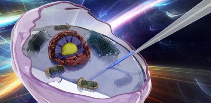
To demonstrate the nanowire probe function, the researchers inserted it into single living HeLa cells, human cancer cells named after Henrietta Lacks, a cancer patient from whom the cell line was derived. The device’s sensitivity to RI enabled the team to observe changes in cell morphology and intracellular microenvironment during a stage of cell development and maturation known as apoptosis. Quantitative detection and analysis of the refractive indices naturally occurring in single living cells during apoptosis can help to advance understanding of cell life events and disease.
This research set a precedent for real-time, in situ sensing and tracking of early cellular apoptosis by using a label-free nano-optical device in living HeLa cells. It will not only contribute to scientific knowledge of cell life events, but also broaden the application scope of fiber nanosensors. At the same time, the strategy of inserting functional nanomodules into single cells overcomes barriers to obtaining information about the interactions between internal and external cell media, lighting a path for new ways to combine the advantages of fiber optics and cell biology. In the future, real-time monitoring of temperature and substance concentration in cells may be realized through the design of different structures, and the evolution of cell physiological processes may be further explored.
Read the open access article by Danran Li et al., “Label-free fiber nanograting sensor for real-time in situ early monitoring of cellular apoptosis,” Adv. Photonics 4(1), 016001 (2022), doi 10.1117/1.AP.4.1.016001.
Journal
Advanced Photonics
DOI
10.1117/1.AP.4.1.016001
Article Title
Label-free fiber nanograting sensor for real-time in situ early monitoring of cellular apoptosis
Article Publication Date
6-Jan-2022


