In a groundbreaking achievement that marks a significant leap in neurotoxicology and structural biology, researchers at Stockholm University have unveiled the molecular architecture of one of the deadliest toxins known to humanity: the botulinum neurotoxin (BoNT). This toxin, regarded as the most potent poison discovered to date, is approximately a million times more toxic than cobra venom. The research, meticulously conducted using advanced cryo-electron microscopy, dissects the entire 14-subunit protein complex enveloping the botulinum toxin, shedding light on its intricate stabilization, delivery, and release mechanisms. These revelations, recently published in Science Advances, not only deepen our understanding of the toxin’s molecular machinery but also herald new avenues for medical innovation and therapeutic intervention.
Botulinum toxin, synthesized by the anaerobic bacterium Clostridium botulinum, is infamously known for inducing botulism – a severe and often fatal paralytic illness. Despite its high toxicity, BoNT paradoxically serves a suite of critical medical applications, including treatments for chronic migraines, dystonia (muscle spasms), hyperhidrosis (excessive sweating), and aesthetic enhancements such as wrinkle reduction. The toxin’s dual nature—lethal yet medicinal—has captivated scientific interest, particularly concerning how it achieves such precise neuromuscular targeting without widespread systemic damage.
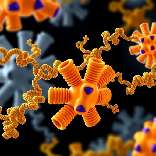
A pivotal aspect of the toxin’s efficacy lies in its natural convoy: a large molecular assembly consisting of 14 distinct protein subunits. This multiprotein complex encapsulates the toxin, providing it with protection against the acidic and proteolytic milieu of the gastrointestinal tract and facilitating its transit across the intestinal barrier into systemic circulation. Professor Pål Stenmark, the study’s lead investigator and a neurochemistry expert at Stockholm University, explains, “The toxin doesn’t act in isolation. It is cloaked within this sophisticated complex that ensures its survival, delivery, and ultimate release at the neuromuscular junction.” Understanding the structural composition of this complex has been a formidable challenge—until now.
The research team employed cryo-electron microscopy (cryo-EM), a Nobel Prize-winning imaging technique renowned for its ability to capture biomolecules in near-native states at near-atomic resolution. This technique involves flash-freezing samples to preserve their native conformations, followed by collection of thousands of two-dimensional images. These are computationally integrated into comprehensive three-dimensional reconstructions that reveal detailed structural insights. Through cryo-EM, the researchers visualized the entire botulinum toxin complex, including its specific arrangement and interactions among subunits.
The resulting molecular blueprint reveals a unique structural anchoring mechanism whereby the toxin itself, depicted in pink in the imagery, nestles atop the complex’s architecture. The surrounding protein subunits collectively form a protective shell, shielding the toxin from environmental stress and enzymatic degradation within the gut. At the core of this complex lies a narrow central pore constituted by the HA70 protein, which serves as the anchoring site for the toxin, stabilizing its conformation while orchestrating its eventual release.
This discovery illuminates the sophisticated strategy BoNT employs to avoid premature neutralization within the host and accomplish targeted delivery. The high-resolution structural data underscore how each protein component contributes synergistically to preserve toxin integrity during intestinal passage and facilitate translocation into the bloodstream. The precise elucidation of this process resolves longstanding questions about the molecular basis of the toxin’s extraordinary stability and potency.
Beyond fundamental biological significance, the elucidation of the botulinum neurotoxin complex’s structure has immense translational potential. With this molecular map in hand, scientists and pharmaceutical developers can now explore novel methodologies to neutralize or inhibit the toxin at multiple junctures in its deployment. Such strategies could dramatically improve countermeasures against botulism, which remains a public health concern globally, particularly in food safety and bioterrorism threat scenarios.
Moreover, the detailed understanding of how the toxin complex travels, stabilizes, and releases its payload can be harnessed to engineer improved therapeutic BoNT derivatives. These refined biologics might offer enhanced specificity, reduced side effects, and expanded applications in neuromuscular disorders or beyond. “Our findings open up avenues for harnessing the toxin’s mechanisms in therapeutic design,” emphasizes Stenmark. “By dissecting how nature’s most dangerous toxin operates on a molecular level, we gain critical insights that can be redirected toward healing.”
The study specifically examined the toxin complex present in NeuroBloc, a pharmaceutical preparation closely related to the widely recognized Botox formulation. This focus offers immediate relevance to clinical practice and drug development, providing a structural framework to optimize existing treatments and innovate next-generation neurotoxin-based therapeutics.
The researchers emphasize that the novel imagery and data do more than satisfy scientific curiosity—they dismantle the opaque barrier that previously limited mechanistic understanding of BoNT. The cryo-EM-derived structural model frames future investigations aimed at dissecting toxin dynamics, interactions with neuronal receptors, and immune evasion tactics. Additionally, it serves as a foundation for rational drug design involving structure-based modifications to modulate toxin activity.
By clarifying the architecture and function of the complete 14-subunit botulinum neurotoxin B complex, the study marks a definitive stride forward in neurotoxin research. The precision of this molecular template promises to guide future endeavors spanning basic biology, medicine, and pharmacology, ultimately balancing the toxin’s menacing power with the potential for therapeutic benefit.
In summary, Stockholm University’s pioneering work elucidates the molecular choreography of botulinum toxin’s assembly and deployment, unveiling a unique central pore anchoring and a protective multi-subunit complex that underpins the toxin’s unmatched lethality and therapeutic versatility. This landmark research not only advances molecular neurochemistry but also galvanizes the scientific community toward innovative therapies combating botulism and enhancing neuromodulation.
Subject of Research: Not applicable
Article Title: Structure of the Complete 14-subunit Botulinum Neurotoxin B Complex Reveals a Unique Anchoring Through the Narrow Central Pore of HA70
News Publication Date: 27-Aug-2025
Web References:
http://dx.doi.org/10.1126/sciadv.adx5058
Image Credits: Pål Stenmark
Keywords: botulinum toxin, neurotoxin, botulism, cryo-electron microscopy, molecular structure, protein complex, neurochemistry, NeuroBloc, HA70, toxin stabilization, therapeutic neurotoxin, neurotoxin delivery
Tags: advancements in structural biologyaesthetic uses of neurotoxinsbotulinum neurotoxin structurechronic migraine treatmentsClostridium botulinum bacteriumcryo-electron microscopy applicationsdual nature of botulinum toxinmedical applications of botulinum toxinneuromuscular targeting mechanismsneurotoxicology research breakthroughsprotein complex architecturetherapeutic interventions for botulism


