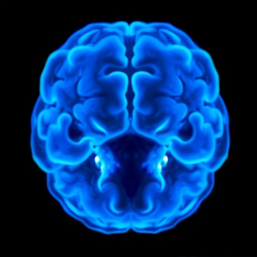

In a groundbreaking advancement in neuroscience, Stanford University researchers have devised a novel technique to achieve transparency in mouse scalp, unlocking possibilities for observing real-time brain development. This remarkable method allows scientists to employ non-invasive imaging techniques to examine neural pathways and their formation in juvenile mice, a crucial stage for understanding brain function and development. As these young mammals undergo significant neurological changes, traditional imaging methods have fallen short, with prior techniques either being too invasive or unable to provide the required clarity.
The innovative approach leverages a compound called ampyrone, which, when rubbed onto the skin, alters the refractive properties of the water within the underlying tissues. This shift in refractive index renders the skin virtually invisible to visible light, effectively eliminating the scattering that typically obscures observations of internal structures. This transformation invites comparisons to viewing objects through fog—prior to treatment, details are indistinct, but post-treatment, clarity is achieved, revealing activities occurring deep within the mouse’s brain.
Earlier methods of imaging the brains of living specimens have often relied on cumbersome surgical techniques that not only harm the animals but also alter their natural behavior. The limitations of these traditional methods have constrained researchers to limited insights, especially regarding how the neural circuits adapt as the organism matures. With the introduction of this new transparency technique, researchers can now repeatedly visualize the same juvenile mouse over days and weeks, monitoring changes in neural pathways as the animal grows, revealing invaluable data regarding brain development.
Guosong Hong, an assistant professor of materials science and engineering and the senior author of the study published in the journal PNAS, expressed his excitement about the potential this method holds for neuroscience research. “This opens a literal window to peek into the brain’s development,” he stated, emphasizing the capability to capture both the structural details of neurons and the dynamic activity within them over time. The benefits of this technique extend not just to observation but also to understanding the mechanisms of neurological disorders, where disrupted development can lead to long-term impacts.
Historically, light scattering has posed significant challenges when imaging through skin and other tissues, as light waves interact with various biological materials, making it difficult to observe internal structures without distortion. By matching the optical properties of the water within tissues to those of biomaterials, the researchers achieved a degree of transparency that was previously unfathomable. Light scattering is diminished as similar refractive indices allow light to pass through without significant deviation, rendering the skin virtually transparent.
The scientists’ approach aligns closely with principles of optics, relying on the interaction of light with matter. Mark Brongersma, a professor and co-author of the study, underscored the significance of applying fundamental physics to biological systems. The clarity achieved not only presents a practical solution to previous imaging barriers but also encourages a deeper exploration into the realms of biology and physics, bridging gaps previously thought to be insurmountable.
The researchers built on prior successful efforts that allowed them to make tissues transparent to red light, opening up discussions about new imaging practices. However, ampyrone’s unique properties enable the use of the entire visible light spectrum, thus enhancing imaging capabilities. In juvenile mice, whose skulls are still developing, this transparency facilitates clearer observations of neural activity linked with fluorescent markers commonly used to track neuronal functions.
Through this advanced technique, researchers have documented significant alterations in neuronal activity in response to external stimuli, providing a comprehensive view of the dynamic changes in the brain’s circuitry as the mice mature. This long-term imaging potential promises significant advances in understanding various neurodevelopmental processes and disorders that can manifest during an organism’s growth, further refining the methodologies employed to track such developments.
As the researchers continue to refine this technique, they anticipate applications extending into broader domains of neuroscience and potentially understanding human neurodevelopmental conditions. Given that the ability to monitor and analyze the developing brain in real-time opens numerous doors, it represents a major leap forward in methodological approaches within the scientific community.
Despite the promising results, further investigation is necessary to validate the long-term safety and effectiveness of this technique in various applications. Researchers are already envisioning future adaptations that could allow them to apply this method to different species or even different biological contexts, providing insights into evolutionary changes across taxa. As they push the boundaries of current scientific practices, the Stanford team’s findings stand as a testament to the convergence of disciplines—physics, biology, and engineering all contributing to a clearer understanding of life’s most intricate processes.
By illuminating the pathways of neuronal connections in adolescence, the research not only enhances scientific understanding but holds potential for clinical applications. Whether through insights into learning behaviors or preemptive measures for neurodevelopmental disorders, the implications of this work may ultimately shape the future landscape of neuroscience, fostering a clearer understanding of the brain’s complexity.
This remarkable study not only lays the groundwork for future innovations in imaging techniques but also raises critical questions about the nature of neural circuitry. With these new tools, researchers are ideally positioned to unlock the mysteries of brain development and pave the way for interventions that could transform our approach to mental health and neurological disorders.
Such progress emphasizes the need for continued exploration and optimization of these imaging techniques, ensuring that they remain non-invasive and accessible for various types of research. Ultimately, this pioneering work exemplifies the thrill of scientific discovery, illustrating how interdisciplinary approaches can yield transformative results that propel human understanding further into the uncharted territories of the brain.
Subject of Research: Neural Imaging in Juvenile Mice
Article Title: Color-neutral and reversible tissue transparency enables longitudinal deep-tissue imaging in live mice
News Publication Date: 26-Aug-2025
Web References: Stanford News
References: Proceedings of the National Academy of Sciences, DOI: 10.1073/pnas.2504264122
Image Credits: The Hong Lab
Keywords
Imaging, Neural Development, Optical Properties, Mouse Model, Neuroscience, Non-invasive Techniques.
Tags: advancements in neuroscience researchampyrone compound in imaginganimal behavior and surgical techniquesclarity in brain imaging methodsjuvenile mouse brain developmentmouse brain imagingneural pathway observationnon-invasive neuroscience techniquesreal-time brain observation methodsrefractive index alteration techniquestraditional imaging limitationstransparency in mouse scalp


