For decades, the understanding of vertebrate spinal cord anatomy rested on a fundamental distinction: tetrapods—vertebrates with four limbs—exhibit pronounced spinal enlargements corresponding to their forelimbs and hind limbs, a neural adaptation supporting complex limb movement. Fish, lacking limbs, were traditionally thought to possess no such spinal enlargements. However, groundbreaking research from Nagoya University in Japan challenges this long-held assumption. This study uncovers the presence of previously undetected spinal enlargements in zebrafish, revealing a significant paradigm shift in our comprehension of vertebrate neuroanatomy and evolutionary biology.
Tetrapods display two distinct spinal enlargements: the cervical enlargement associated with nerve supply to the forelimbs, and the lumbar enlargement linked with the hind limbs. These enlargements accommodate dense clusters of motor neurons responsible for intricate musculature and sensory feedback essential for limb coordination. In contrast, fish species, devoid of limbs and instead equipped with fins, were presumed to have uniform spinal cords without any specialized swellings. This perspective overlooked the potential neuroanatomical complexity associated with paired and unpaired fins in fish.
Naoyuki Yamamoto and his team hypothesized that due to zebrafish possessing paired pectoral and pelvic fins—analogous to the forelimbs and hind limbs in tetrapods—there might be corresponding spinal cord enlargements. To test this hypothesis, they embarked on a meticulous investigation combining advanced histological techniques and three-dimensional tissue visualization methods, pushing beyond the limitations of conventional anatomical scrutiny.
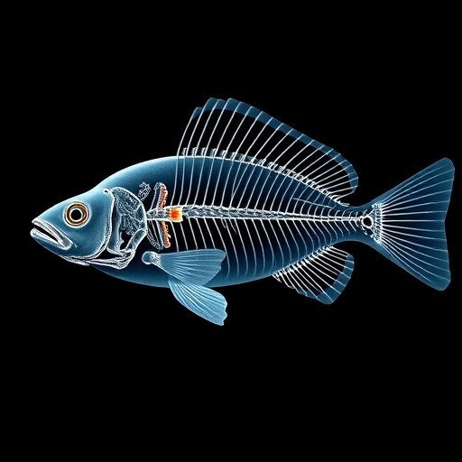
Identifying the spinal cord regions responsible for innervating various zebrafish fins required precise mapping. While prior studies had elucidated innervation patterns for pectoral, dorsal, and caudal fins, the researchers concentrated on less understood pelvic and anal fins. Employing immunohistochemistry—a method that tags neuron cell bodies and axons with fluorescent markers—they stained entire zebrafish specimens to trace complex neural pathways. Further, the team utilized a modified CUBIC technique, a cutting-edge tissue-clearing protocol, to render the specimen optically transparent, thereby enabling deep imaging of spinal nerve structures without physical dissection.
Serial sections of the spinal cord allowed the scientists to quantify changes in cross-sectional areas of both spinal cord tissue and gray matter with unprecedented accuracy. The analysis yielded striking results: not only did zebrafish exhibit spinal enlargements associated with paired fins, but there were also subtle yet definitive enlargements connected to unpaired fins—dorsal, anal, and caudal. These findings demonstrate that the zebrafish spinal cord, subtle as it may be, displays region-specific hypertrophy akin to the spinal enlargements well documented in tetrapods.
This discovery carries profound implications for evolutionary biology. The presence of spinal enlargements in fish suggests that these neuroanatomical features predate the evolution of terrestrial limbs and may have originally evolved to support locomotion mediated by paired and unpaired fins. The traditional view that spinal enlargements are exclusive adaptations for limbs is thus incomplete. Instead, these structures likely represent a more ancient neural adaptation for controlling complex appendages, whether fins or limbs.
The research further illuminates the evolutionary trajectory of vertebrates transitioning from aquatic to terrestrial environments. Tetrapods evolved from finned ancestors, but only paired fins persisted and transformed into limbs, while unpaired fins largely disappeared. The corresponding spinal enlargements for paired appendages were retained and possibly elaborated upon to meet the demands of life on land. The nuanced spinal enlargements associated with unpaired fins in zebrafish challenge the neat dichotomy between fish and tetrapod spinal anatomy, suggesting a continuum of neural specialization aligned with the type and function of appendages.
This innovative study also underscores the value of integrating modern tissue-clearing techniques with traditional histology to reveal subtle anatomical features invisible under routine observation. The methodology—combining immunohistochemical labeling with CUBIC clearing—opens new avenues for neuroanatomical research across species and organ systems, enabling researchers to peer deep into opaque tissues with cellular resolution and spatial context.
Moreover, the findings could inspire reassessment of neurological evolution and developmental biology. Understanding how spinal enlargements develop in fish may shed light on genetic and molecular mechanisms regulating neural circuit formation for motor control. The study highlights potential conserved pathways underlying appendage innervation, bridging gaps in knowledge between piscine and tetrapod neurodevelopment.
This breakthrough prompts a reexamination of neurofunctional specialization, raising questions about the extent to which spinal enlargements correlate with fine motor control or sensory processing in fins. Since fins engage in complex swimming maneuvers, balance, and substrate interaction, the modest enlargements observed may reflect adaptations optimized for aquatic locomotion dynamics rather than terrestrial weight-bearing or manipulation.
In summary, zebrafish possess spinal cord enlargements associated with all fin types—paired and unpaired—though subtly expressed and requiring advanced histological techniques for detection. This discovery challenges existing paradigms, suggesting that spinal enlargements are an evolutionary conserved neuroanatomical feature linked to appendage innervation predating the emergence of limbs. It enriches our understanding of vertebrate neural evolution and enhances the conceptual framework encompassing motor system adaptations across aquatic and terrestrial contexts.
The elucidation of spinal enlargements in zebrafish invites broader exploration of spinal neuroanatomy across diverse fish species, potentially uncovering iterative or divergent patterns of neural specialization corresponding to ecological niches and locomotor strategies. Such research has the potential to reconstruct a more detailed evolutionary map of vertebrate motor systems and inform biomedical approaches to spinal cord injury and regeneration by revealing fundamental principles of spinal cord organization.
Professor Yamamoto’s work represents a milestone in neuroevolutionary research, as published in the journal Brain, Behavior and Evolution, setting the stage for future inquiries into the intersection of anatomy, function, and evolutionary history. The meticulous application of advanced imaging and histological techniques underscores the importance of technological innovation in revising long-standing biological assumptions and deepening our understanding of complex biological systems.
Subject of Research: Animals
Article Title: Identification of “spinal enlargements” correlating with paired and unpaired fins in zebrafish
News Publication Date: 29-Aug-2025
Web References: http://dx.doi.org/10.1159/000548184
References: Yamamoto, N., Takaoka, R., & Hagio, H. (2025). Identification of “spinal enlargements” correlating with paired and unpaired fins in zebrafish. Brain, Behavior and Evolution. https://doi.org/10.1159/000548184
Image Credits: Naoyuki Yamamoto
Keywords: Life sciences, Evolutionary biology, Evolutionary theories, Ecological adaptation, Neuroethology
Tags: evolutionary biology of fishfins vs limbs in evolutiongroundbreaking spinal cord discoverieslimb coordination in vertebratesmotor neuron clusters in fishNagoya University research findingsneuroanatomical adaptations in aquatic speciesspinal cord anatomy in fishspinal enlargements in vertebratestetrapod neuroanatomy comparisonvertebrate evolution and neurobiologyzebrafish spinal cord research


