Administering the hypoxia-inducible factor prolyl hydroxylase inhibitor roxadustat ameliorated femoral trabecular bone microarchitectural deterioration in an established rat model of chronic kidney disease-mineral and bone disorder, according to laboratory findings reported by researchers at Qinghai University and collaborating institutions. The experimental evaluation demonstrated that roxadustat preserved trabecular network density, restored bone volume fraction, and elevated bone mineral density over an eight-week oral intervention. Crucially, the observed skeletal protection did not occur in isolation from circulating conditions; rather, the preservation of femoral structural integrity was documented synchronously with the downregulation of circulating and tissue fibroblast growth factor 23, the suppression of systemic micro-inflammatory markers, and the molecular derepression of osteogenic Wnt and beta-catenin signaling cascades in femoral bone tissue.
Chronic kidney disease-mineral and bone disorder, commonly designated CKD-MBD, represents a complex systemic condition linked to substantial morbidity and mortality in individuals experiencing progressive renal dysfunction. Under recent clinical framework revisions, including the 2025 KDIGO Controversies Conference report, the syndrome has been conceptualized around two major systemic manifestations: CKD-associated cardiovascular disease and CKD-associated osteoporosis. The skeletal phenotype of this condition features progressive skeletal mass loss, architectural disruption of trabecular lattices, and impaired structural quality, which together drive elevated fracture risks. Conventional clinical management strategies have historically centered around correcting downstream serum calcium and phosphate disturbances and managing secondary hyperparathyroidism; however, these conventional therapeutic approaches often demonstrate limited efficacy in halting progressive structural bone loss. Moreover, accumulating biomedical evidence suggests that persistent systemic micro-inflammation and disrupted iron homeostasis act as primary drivers of aberrant fibroblast growth factor 23 secretion, linking renal dysfunction directly to impaired skeletal remodeling.
Fibroblast growth factor 23, abbreviated FGF23, functions as a principal humoral mediator produced predominantly by osteocytes within the skeletal compartment. Under physiological conditions, FGF23 regulates systemic mineral balance, yet in advanced renal failure, it accumulates pathologically. Elevated hepatic hepcidin production, stimulated by inflammatory markers such as C-reactive protein, restricts physiological iron utilization and precipitates functional iron deficiency. This combined inflammatory and iron-restricted environment acts as a potent stimulus for aberrant osteocytic FGF23 synthesis and secretion. Once overexpressed, FGF23 engages its local receptor FGFR1 within bone tissue, triggering the upregulation of Dickkopf-1, or DKK1, a potent endogenous antagonist of canonical Wnt signaling. DKK1 binding blocks the nuclear translocation of beta-catenin and promotes its proteasomal degradation, thereby quenching canonical osteogenic transcriptional activity, suppressing osteoblast differentiation, and destabilizing the dynamic cellular balance required to maintain trabecular integrity.
To investigate whether roxadustat could alter this systemic-local pathological axis beyond its standard role in managing renal anemia, the research team employed specific-pathogen-free male Sprague-Dawley rats weighing between 180 and 220 grams. Animals were housed in barrier facility environments under controlled temperature, humidity, and twelve-hour light cycles with ad libitum food and water access. The CKD-MBD pathology was induced through a surgical and toxicological protocol consisting of a left unilateral nephrectomy performed under general isoflurane anesthesia, followed by daily oral gavage of adenine at a dose of 150 milligrams per kilogram for four consecutive weeks. Adenine gavage produced extensive tubulointerstitial damage, marked elevation of serum creatinine and blood urea nitrogen, and severe trabecular bone loss. A sham-operated group underwent identical abdominal incisions and kidney exposure without nephrectomy or adenine feeding to serve as a surgical reference.
Following the initial four-week modeling period, animals with confirmed CKD-MBD were randomized into three experimental arms alongside the sham control group, with six animals assigned to each cohort. The treatment arms consisted of an untreated CKD-MBD model control group receiving one percent sodium carboxymethyl cellulose vehicle, a low-dose roxadustat group receiving 50 milligrams per kilogram, and a high-dose roxadustat group receiving 100 milligrams per kilogram. Because adenine withdrawal can permit spontaneous renal regeneration, a maintenance dose of adenine at 150 milligrams per kilogram was administered every three days across all disease groups throughout the subsequent eight-week treatment phase. Roxadustat and vehicle suspensions were delivered by oral gavage three times weekly for eight consecutive weeks, yielding a total experimental study timeline of twelve weeks before final physiological and structural tissue harvesting.
Hematological and biochemical analyses demonstrated marked systemic improvements following roxadustat intervention. The adenine model group exhibited severe anemia, marked by sharp reductions in red blood cell count, hemoglobin, and hematocrit, all showing statistical significance at p values below 0.001 compared to sham controls. Roxadustat treatment dose-dependently reversed these deficits; notably, hemoglobin and red blood cell counts in the high-dose group exceeded sham levels, reflecting known pro-erythropoietic responses driven by uninhibited erythropoietin gene induction. Renal functional biomarkers also shifted substantially: serum blood urea nitrogen and creatinine, which rose 13.5-fold and 8.5-fold respectively in untreated disease models, were significantly attenuated by low-dose roxadustat and reduced even further in the high-dose cohort. In parallel, systemic mineral derangements were mitigated: roxadustat significantly lowered elevated intact parathyroid hormone, serum phosphorus, and alkaline phosphatase, while increasing circulating calcium and 1,25-dihydroxyvitamin D3 concentrations.
Renal histopathology confirmed structural preservation in the renal parenchyma that mirrored the serological improvements. Hematoxylin and eosin staining of kidney sections from the untreated model group displayed widespread structural damage, including severe tubular dilation, epithelial brush-border loss, extensive vacuolar degeneration, interstitial inflammatory infiltrates, and tubular lumen crystal deposits. Both the tubular injury score and the tubular dilation score were significantly decreased in animals receiving roxadustat at 50 milligrams per kilogram, with the 100 milligram per kilogram dose producing additional histological protection. Complementary Masson’s trichrome staining showed heavy collagen fiber deposition in the renal interstitium of model animals; quantitative image analysis demonstrated that roxadustat significantly reduced this collagen volume fraction in a dose-dependent manner, confirming an attenuation of tubulointerstitial fibrosis.
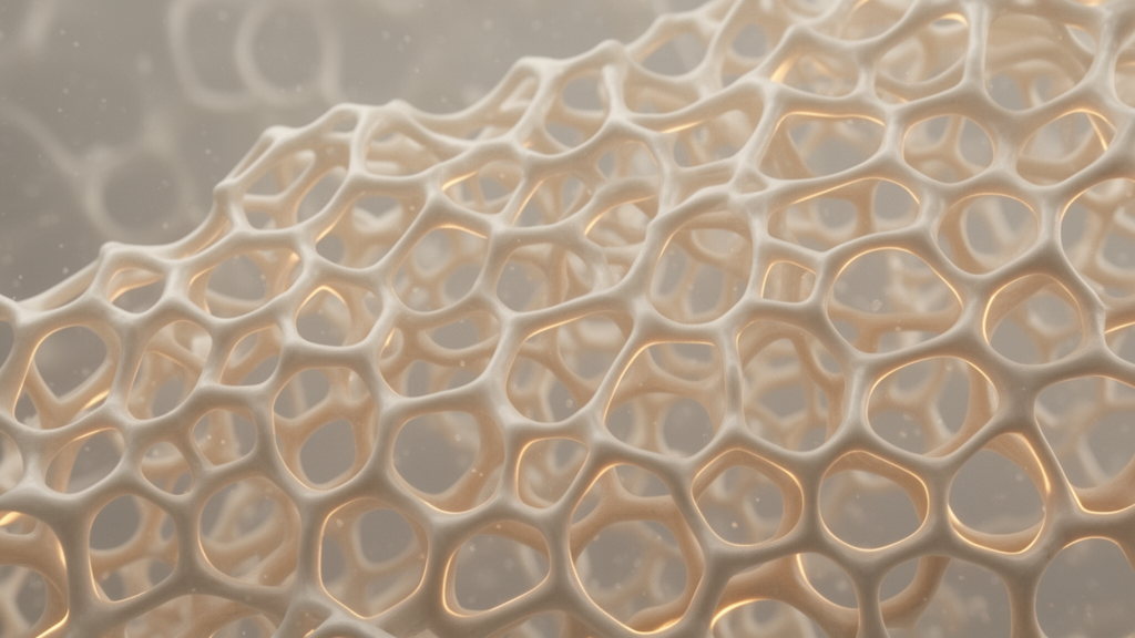
High-resolution micro-computed tomography scans of the distal femoral metaphysis revealed substantial preservation of trabecular architecture in roxadustat-treated rats compared to untreated model controls. While model animals exhibited sparse, thinned, and fractured trabeculae with severed structural connectivity, animals receiving roxadustat maintained continuous, dense trabecular networks. Quantitative morphometric calculations confirmed that bone mineral density, bone volume fraction, and trabecular connectivity density, which dropped significantly in model rats, were substantially elevated by roxadustat therapy. In trabecular geometric dimensions, roxadustat treatment significantly increased trabecular number and decreased trabecular separation. While the lower dose of 50 milligrams per kilogram did not achieve a statistically significant improvement in trabecular thickness alone, the 100 milligram per kilogram regimen significantly elevated trabecular thickness compared to the model group, demonstrating structural restoration across microarchitectural parameters.
Molecular profiling provided clear evidence of coupled changes across systemic humoral mediators and local skeletal signaling proteins. Enzyme-linked immunosorbent assays demonstrated that elevated circulating C-reactive protein, hepcidin, and intact FGF23 in model rats were markedly suppressed by roxadustat administration in a dose-dependent fashion. Western blot assays of harvested bone and kidney homogenates similarly confirmed significant reductions in tissue FGF23 abundance. In femoral bone extracts, western blotting revealed that untreated disease animals exhibited marked upregulations of FGFR1 and the Wnt inhibitor DKK1 alongside diminished beta-catenin levels; roxadustat treatment suppressed FGFR1 and DKK1 expression while simultaneously restoring beta-catenin protein concentrations. Immunohistochemical evaluations corroborated these protein patterns, verifying decreased intraosseous DKK1 and FGF23 staining density alongside restored cytoplasmic and nuclear beta-catenin localization in osteocytes and osteoblasts.
Statistical evaluation through pooled Pearson correlation analysis of all 24 experimental animals revealed that femoral bone mineral density correlated significantly and negatively with serum FGF23, intact parathyroid hormone, phosphorus, blood urea nitrogen, creatinine, C-reactive protein, and hepcidin, while correlating positively with serum calcium. In contrast, bone mineral density did not exhibit a statistically significant correlation with circulating hemoglobin levels, demonstrating that the structural bone preservation documented in this model was not merely a secondary byproduct of anemia correction. The synchronous alignment of reduced inflammatory markers, lower circulating and skeletal FGF23, decreased DKK1, and increased beta-catenin abundance indicates that roxadustat acts across a coordinated systemic-local pathway involving micro-environmental metabolic regulation and skeletal Wnt signaling preservation.
The study authors highlighted several methodological limitations that restrict the broader interpretation of their experimental findings. Because the holistic in vivo animal model intervened systemically, the study design could not strictly uncouple the direct osteogenic actions of skeletal hypoxia-inducible factor activation from the indirect systemic benefits of improved renal filtration and reduced systemic inflammation. Furthermore, dynamic histomorphometry, cortical bone microstructural parameters, mechanical bone strength testing, and vascular calcification endpoints were not evaluated. The research team also noted the absence of a healthy rat control receiving high-dose roxadustat alone to profile drug effects in the absence of renal pathology. Consequently, while these preclinical observations demonstrate that roxadustat counters bone microarchitectural deterioration in adenine-nephrectomy rats, clinical extrapolation to human chronic kidney disease-associated osteoporosis requires targeted clinical trials.
Subject of Research: Pharmacological preservation of femoral trabecular microarchitecture by roxadustat in adenine-induced chronic kidney disease rats
Article Title: Roxadustat ameliorates bone microarchitectural deterioration in CKD-MBD rats in association with mitigated systemic micro-inflammation and restored FGF23–Wnt/β-catenin signaling
Article References: Shan, Z., Liu, Y., Sun, X., Li, Z., Ma, J., Wang, Y., Ai, S., Yang, Y., & Luo, P. (2026). Roxadustat ameliorates bone microarchitectural deterioration in CKD-MBD rats in association with mitigated systemic micro-inflammation and restored FGF23–Wnt/β-catenin signaling. Frontiers in Pharmacology, 17, Article 1957451. https://doi.org/10.3389/fphar.2026.1957451
Image Credits: AI Generated
DOI: 10.3389/fphar.2026.1957451
Keywords: roxadustat, CKD-MBD, bone microarchitecture, FGF23, Wnt beta-catenin signaling, DKK1, chronic kidney disease, hepcidin, C-reactive protein, secondary hyperparathyroidism, micro-CT, adenine model, nephrectomy, trabecular bone, FGFR1, hypoxia-inducible factor prolyl hydroxylase inhibitor
News Source: Jerry Hayes. (November 17, 2026). Roxadustat Protects Bone Microarchitecture in Chronic Kidney Disease Rat Model. Scienmag.