A retrospective radiological investigation tracking robotic-arm-assisted cementless total knee arthroplasty has identified distinct temporal milestones in biological bone integration, demonstrating that newly formed trabecular bone bridges develop in over ninety percent of knees during the initial postoperative months. Historically, the gold standard for total knee arthroplasty fixation was cemented fixation. However, concerns regarding long-term failure rates and aseptic loosening accompanied by failure of the bone-cement interface—particularly among young obese patients—have spurred widespread interest in cementless alternatives that achieve biological fixation. The retrospective study analyzed two hundred twenty-one consecutive patients representing three hundred one knees treated for primary osteoarthritis using robotic navigation and a modern porous knee system, revealing that structural trabecular lines emerged in over ninety percent of treated joints at a mean interval of four and a half months following surgical implantation.
Biologic fixation requires an orchestrated cellular cascade wherein host bone actively migrates into and integrates with the porous scaffolding of prosthetic hardware. Unlike bone ongrowth, which typically relies on textured surface alterations such as grit blasting or plasma spraying to facilitate superficial apposition, true bone ingrowth depends heavily upon specialized metal porosity with pore dimensions optimized between fifty and four hundred micrometers to allow osseous penetration and vascularization. To assess the viability of using routine standard plain radiographs as an accessible alternative to highly specialized techniques like radiostereometric analysis or artifact-reduction computed tomography, two independent orthopedic investigators systematically tracked the progression of primary radiographic markers across serial examinations. The investigators specifically evaluated the incidence and timing of new trabecular bone line formation, the emergence and subsequent resolution of immediate radiolucent gaps along component borders, and the secondary appearance of focal bone resorption zones across follow-up appointments extending to a mean of forty-six and a half months.
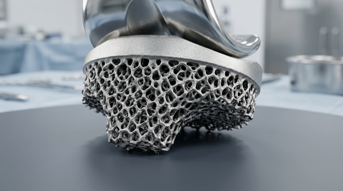
All surgical procedures within the evaluated cohort were conducted across a single academic institution by six specialized orthopedic surgeons employing the Mako Robotic-Arm-Assisted Surgery System. The robotic platform facilitated precise bone resections based on patient-specific preoperative three-dimensional computed tomography segmentation and dynamic soft-tissue balancing. Every patient received the Triathlon Tritanium cementless knee implant system, featuring a three-dimensional printed titanium-coated tibial tray, a cobalt-chromium-coated femoral component, and an advanced cross-linked polyethylene articular insert. Surgeons adhered to a restricted kinematic alignment philosophy aimed at minimizing collateral ligament releases by preserving individual constitutional joint anatomy within defined boundaries, targeting component placement within four degrees of neutral and limb alignment not exceeding six degrees of varus. Postoperative care adhered strictly to a standardized active rehabilitation protocol that introduced immediate full weight-bearing ambulation alongside active and passive mobilization on the initial day of surgery.
Standardized weight-bearing anteroposterior and true lateral radiographs obtained on postoperative day one and at regular intervals spanning up to seventy-one months revealed that nine out of ten knees established visible new trabecular bone formation. New trabeculae appeared most densely localized in regions undergoing high mechanical load transfer, specifically along the medial aspects of the tibial keel where sixty-seven point two percent of knees exhibited trabecular development, as well as the lateral keel in thirty-six point nine percent of cases. Lateral views similarly demonstrated frequent trabecular bridging beneath the posterior tibial tray in twenty-three point six percent and along the posterior keel in thirty-six point two percent of knees. Conversely, new trabeculae appeared along the femoral component in only seventeen point nine percent of evaluated joints.
In contrast to the constructive bridging represented by trabecular lines, early radiolucent lines manifested immediately following surgery in thirty-three point six percent of the total knee cohort, representing one hundred one distinct knees. These thin radiolucent intervals at the bone-prosthesis boundary occurred most frequently across the femoral anterior chamfer region, affecting thirteen point four percent of knees, while the tibial interface displayed radiolucencies primarily beneath the posterior tray in five point six percent of cases. Rather than heralding mechanical loosening or fixation failure, serial follow-up demonstrated that ninety-six percent of these initial radiolucent gaps resolved spontaneously over time, with complete radiographic disappearance recorded in ninety-seven of the one hundred one affected joints. Investigators observed that radiolucent line obliteration occurred at an average postoperative interval of eight point nine months, with the radiographic disappearance of the gap systematically preceded by adjacent trabecular bone bridging.
Focal zones of bone resorption, categorized operationally as diffuse areas of reduced radiographic bone mineral density rather than discrete linear interfaces, developed in forty-seven point five percent of the overall cohort. Resorption patterns emerged predominantly within the femoral anterior chamfer region in forty-one point two percent of knees, as well as beneath the medial tibial tray in thirty-six point five percent and the anterior tibial tray in thirty-six point nine percent of knees. Biomechanical principles, including those described by Wolff’s law, provide a framework for interpreting these localized changes, wherein structural bone adapts dynamically to altered stress distributions. As newly established trabecular formations around the central tibial keel absorb increased proportions of compressive and shear forces, neighboring peripheral bone zones experience relative unloading, culminating in localized proximal stress shielding and subsequent cortical and cancellous resorption.
Statistical evaluation of the timing of these radiographic events uncovered a defined chronological sequence governing cementless implant integration. Overall, new trabecular bone bridging emerged early in the recovery timeline at an average of four point five months, whereas focal stress-shielding resorption presented significantly later, requiring an average of sixteen point two months to become radiographically conspicuous. A distinct temporal divergence emerged when comparing joints with and without early radiolucent lines. In knees lacking initial radiolucency, trabecular formation appeared earlier at a mean of four point zero months, compared to five point five months in knees possessing initial gaps. Logistic regression demonstrated that early trabecular formation within five point five months was a strong predictor of subsequent radiolucent gap resolution within eight point nine months, generating an odds ratio of three point nine three.
Exploratory univariate and multivariate logistic regression analyses were conducted to evaluate whether host systemic factors and patient demographic parameters influenced biological fixation or remodeling patterns. Univariate testing initially suggested that elevated body mass index and preexisting hypertension were associated with reduced rates of focal bone resorption, but the hypertensive correlation ceased to achieve statistical significance following comprehensive multivariate adjustment. After adjusting for potential confounding covariates, advancing patient age emerged as the sole statistically significant predictor of trabecular line formation, yielding an odds ratio of one point one zero six. The authors emphasized that this unexpected age association likely represents unmeasured confounding related to reduced baseline bone mineral density, which may render newly formed trabeculae more visually conspicuous against osteopenic backgrounds rather than signifying enhanced cellular integration.
Mid-term mechanical stability and clinical survivorship remained exceptionally high across the entire study period, with radiographic component alignment displaying remarkable consistency from initial postoperative imaging to final follow-up. Measurements evaluating the medial proximal tibial angle, lateral distal femoral angle, and posterior tibial slope revealed no statistically significant deviations over the minimum two-year evaluation window. One patient underwent isolated femoral component revision, while another knee underwent isolated polyethylene liner exchange to address symptomatic ligamentous laxity and joint instability without radiological implant detachment, and no deep prosthetic joint infections were documented.
Despite these favorable findings, several methodological constraints qualify the broader interpretation of the radiographic outcomes. The investigation was conducted as a single-center retrospective analysis utilizing a single robotic surgical platform paired exclusively with a proprietary three-dimensional printed titanium porous knee system, limiting immediate generalizability to alternative implant geometries, coatings, or conventional manual instrumentation. Preoperative bone mineral density screening via dual-energy X-ray absorptiometry was not routinely administered, precluding precise quantification of baseline osteopenia or systemic osteoporosis across aging participants. Furthermore, patient-reported outcome measures were not gathered alongside imaging data, meaning that visual bone ingrowth and regional stress shielding could not be correlated directly with functional knee scores, residual postoperative pain, or subjective patient satisfaction metrics.
The study also faced analytical constraints arising from treating each operated knee as an independent data point without adjusting for intra-patient correlation among individuals undergoing bilateral procedures, which may alter variance calculations. Plain radiography, while widely accessible, cost-effective, and reproducible across standardized centers, inherently lacks the microscopic precision of radiostereometric marker analysis or advanced volumetric computed tomography. Nevertheless, the demonstration that non-progressive early radiolucent lines resolve following initial trabecular bone formation provides clinicians with practical benchmarks for routine postoperative surveillance.
Subject of Research: Radiographic evidence and timeline of biological bone ingrowth following robotic cementless total knee arthroplasty
Article Title: Radiographic features of bone ingrowth in highly porous cementless total knee arthroplasty with minimum 2-year follow-up
Article References: Chan, K. C. A., Cheung, A., Luk, M. H., Leung, K. C. T., Lau, C. M. L., Chang, X., Chiu, K. Y., & Fu, H. (2026). Radiographic features of bone ingrowth in highly porous cementless total knee arthroplasty with minimum 2-year follow-up. Arthroplasty, 8(1), Article 60. https://doi.org/10.1186/s42836-026-00422-6
Image Credits: AI Generated
DOI: 10.1186/s42836-026-00422-6
Keywords: total knee arthroplasty, cementless TKA, bone ingrowth, osteointegration, trabecular line formation, radiolucent lines, bone resorption, stress shielding, robotic-assisted surgery, orthopedic implants, tibial keel, anterior chamfer, biological fixation, radiographic evaluation, knee osteoarthritis, titanium coating, aseptic loosening
News Source: Ophelia Keating. (November 14, 2026). Robotic Cementless Knee Implants Achieve Strong Bone Ingrowth in Retrospective Study. Scienmag.