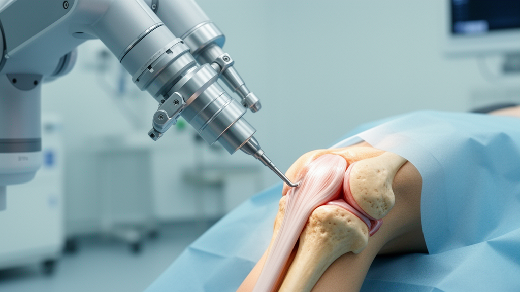
Conventional manual total knee arthroplasty introduces an unintended mechanical phenomenon known as iatrogenic coronal-sagittal coupling, where deviations during surgical cuts in the frontal plane inadvertently skew the sagittal alignment of the reconstructed joint. In a retrospective radiographic cohort study evaluating three hundred and sixty patients, orthopedic researchers identified that an inherent twelve-point-four-degree rotational mismatch between manual tibial instrumentation and the true anatomic placement axis directly converts coronal cutting errors into significant sagittal slope inaccuracies. By contrasting traditional extramedullary manual techniques with computerized robotic systems, the investigation demonstrated that robotic-assisted arthroplasty completely decouples these two planes, neutralizing unintended three-dimensional error transfer and maintaining key functional knee geometric profiles.
Achieving physiological alignment across both the coronal and sagittal planes is fundamental to long-term implant survival, joint stability, and postoperative patient mobility following knee reconstruction. While personalized alignment frameworks such as the Coronal Plane Alignment of the Knee classification have advanced coronal positioning, surgical preservation of sagittal geometry has historically presented substantial clinical hurdles. In the native knee joint, natural coronal parameters and sagittal parameters function with complete anatomic independence, exhibiting no structural correlation. Prior to this investigation, however, surgical researchers had not systematically quantified whether traditional manual cutting guides preserve this baseline anatomical independence during actual bone resection and prosthetic component seating.
The study cohort comprised three hundred and sixty consecutive patients undergoing primary total knee arthroplasty between October 2023 and October 2025 at China-Japan Friendship Hospital, including one hundred and ninety-nine individuals treated with conventional manual instrumentation and one hundred and sixty-one managed with image-based robotic assistance. Preoperative baseline characteristics, including age, biological sex, body mass index, and baseline deformity measurements, exhibited no statistically significant differences between the two surgical groups. Patients enrolled had verified primary knee osteoarthritis classified as Kellgren-Lawrence grade three or four accompanied by significant varus or valgus deformities. Detailed radiological assessments were conducted via standardized standing full-length lower extremity radiographs and lateral knee views, evaluated by two independent blinded orthopedic surgeons.
To evaluate operative deviations in the frontal plane, researchers defined the Tibial Cutting Guide Varus/Valgus Angle, calculated as ninety degrees minus the postoperative medial proximal tibial angle. In the manual cohort, surgeons relied on conventional extramedullary alignment rods aimed proximally at the medial third of the tibial tuberosity and distally toward a reference point three to five millimeters medial to the ankle center. Across the manual group, the mean execution deviation measured zero-point-thirty-four degrees with a standard deviation of two-point-sixteen degrees, capturing the inherent variability of physical extramedullary positioning. In contrast, robotic-assisted procedures integrated preoperative computed tomography mapping with dynamic intraoperative tracking, eliminating guide tilt at the point of origin.
Analysis of postoperative radiographic parameters revealed a distinct cross-planar interaction restricted exclusively to the manual arthroplasty group. Among manual patients, changes in the medial proximal tibial angle demonstrated a statistically significant negative correlation with alterations in the posterior tibial slope, exhibiting a correlation coefficient of minus zero-point-two-hundred-and-nine. When adjusted for patient age, sex, operative laterality, and baseline alignment metrics, this association remained statistically robust. Conversely, the robotic-assisted cohort displayed no correlation between coronal adjustments and sagittal slope modifications, demonstrating that computational navigation prevents coronal plane resections from altering sagittal slope outcomes.
Statistical modeling established that the tibial cutting guide angle served as an independent predictor of postoperative posterior tibial slope in manual procedures, yielding a regression coefficient of zero-point-two-hundred-and-nineteen. Applying stereometric projection principles, this linear coefficient translates into the tangent of an underlying angular offset, indicating an equivalent rotational mismatch of approximately twelve-point-four degrees between the extramedullary guide alignment axis and the surgical prosthesis reference established along the Akagi line. When coronal guide tilt occurs alongside this rotational offset, geometric projection rotates the planned osteotomy, forcing unintended shifts in posterior tibial slope that directly compromise joint biomechanics.
The study also revealed that patients presenting with severe preoperative varus deformities were significantly more vulnerable to this mechanical cascading error. Greater baseline deformities are anatomically coupled with tibial shaft bowing, altered lateral positioning of the tibial tuberosity, and extensive medial plateau bone sclerosis. These combined pathological variations complicate extramedullary guide rod alignment and physical pin fixation, predisposing the manual cutting block to coronal tilt. The resulting tilt is subsequently projected across the twelve-point-four-degree rotational mismatch, culminating in disproportionate sagittal slope deviations in the patients who require the most precise surgical correction.
Preservation of Combined Flexion, defined as the sum of distal femoral flexion and posterior tibial slope, differed markedly between surgical cohorts. Maintaining Combined Flexion within four degrees of baseline anatomy is an established threshold associated with meaningful functional recovery and physiological soft-tissue tension. In the robotic-assisted group, sixty-point-nine percent of patients successfully preserved native Combined Flexion, compared to just twenty-two-point-six percent of patients undergoing manual surgery. Robotic planning actively coordinated sagittal mechanics: when posterior tibial slope was reduced to balance the flexion gap, the robotic system introduced compensatory distal femoral flexion, preserving overall sagittal balance through reciprocal multi-component adjustments.
Deviations in the posterior tibial slope generate well-documented clinical complications in joint reconstruction, including unbalanced flexion gaps, posterior cruciate ligament tension abnormalities, and accelerated polyethylene insert wear. While manual surgeons can implement refined positioning protocols, fluoroscopic checks, or accelerometer-based navigation to diminish cutting guide tilt, physical instrumentation cannot eliminate the twelve-point-four-degree rotational discrepancy inherent to extramedullary referencing. Robotic three-dimensional coordinate registration bypasses anatomical surface proxies, maintaining planar independence and insulating sagittal resection depth from rotational discrepancies.
The authors recognized several methodological limitations inherent to the investigation, including its retrospective non-randomized single-center design and the absence of multi-year clinical functional outcome scores linking Combined Flexion preservation directly to patient-reported satisfaction. Additionally, the manual cohort exclusively followed mechanical alignment principles using manual guides, whereas the robotic cohort utilized a functional alignment philosophy on a single image-based robotic platform, partially confounding instrumentation differences with surgical alignment philosophies. Despite these constraints, the study provides quantified radiographic evidence of three-dimensional coupling in manual arthroplasty and underscores the mechanical mechanism through which robotic guidance achieves planar decoupling.
Beyond the final 360 participants, the trial initially enrolled 408 consecutive patients before applying defined structural exclusions. Patients were excluded for secondary osteoarthritis, previous ipsilateral fracture surgery, severe extra-articular deformities, prior contralateral arthroplasty, or lateral radiographs lacking sufficient femoral condyle overlap. Furthermore, specific anatomical presentations strictly mandated conventional manual procedures. These complex presentations included uncorrectable flexion contractures exceeding 20 degrees under anesthesia, severe tibial plateau bone defects requiring augments or stem extensions, and collateral ligament insufficiency requiring constrained prostheses. To ensure rigorous evaluation, two blinded orthopedic surgeons measured coronal parameters using Matlab alongside lateral views analyzed through RadiAnt DICOM Viewer and ImageJ. Observers repeated measurements across a two-week interval, achieving intraclass correlation coefficients exceeding 0.85. When interobserver discrepancies exceeded three degrees, a third senior surgeon adjudicated to reach consensus.
Operative workflows also incorporated divergent bone resection and implant positioning protocols. For manual reconstructions, femoral preparation required entering the medullary canal one centimeter anterior to the posterior cruciate ligament insertion and beneath the anterior cortex, seating an intramedullary guide fixed at six degrees of valgus. Rotational orientation was aligned to the transepicondylar axis using a four-in-one cutting block set to three degrees of external rotation, while tibial resection targeted a posterior slope of three degrees for posterior-stabilized designs or five degrees for cruciate-retaining implants. In contrast, the robotic group utilized computed tomography data within a functional alignment framework, dynamically tailoring resection depths to soft-tissue tension curves. Sagittal planning permitted femoral component flexion up to seven degrees to match the anterior femoral bow while strictly preventing hyperextension, establishing target slopes of three degrees for posterior-stabilized and five degrees for cruciate-retaining prostheses within rigid predefined planning boundaries.
The research also established an accessible radiographic method for assessing preoperative distal femoral flexion directly on standard lateral radiographs via the distal femoral anatomical axis. Although previous anatomical literature identified a two-degree divergence between the femoral anatomical and mechanical axes, evaluating relative changes before and after surgery effectively canceled out this baseline offset. Applying this measurement revealed unexpected sagittal dynamics across cohorts. Postoperatively, posterior tibial slope decreased in both arms, but the robotic cohort experienced a significantly greater reduction of 6.78 degrees compared to 5.66 degrees in manual knees. However, because robotic navigation concurrently increased distal femoral flexion by 2.34 degrees compared to a 0.17-degree decrease in manual cases, the robotic cohort exhibited a significantly smaller net reduction in Combined Flexion of 4.47 degrees versus 5.83 degrees, demonstrating effective sagittal coordination.
Coronal alignment preservation further differentiated surgical performance when evaluated through the Coronal Plane Alignment of the Knee framework. The robotic cohort achieved significantly superior preservation rates for both arithmetic hip-knee-ankle alignment, at 69.6 percent versus 58.8 percent, and joint line obliquity, at 23.6 percent versus 14.1 percent. Composite parameter analysis in the manual cohort confirmed that joint line obliquity changes were inversely correlated with Combined Flexion alterations, while overall limb alignment shifts showed no such association. Crucially, stereometric modeling illustrated that rotational mismatch in isolation produces negligible slope distortion when cutting guides remain strictly neutral; with zero guide tilt, the postoperative slope equals the planned angle multiplied by the cosine of 12.4 degrees, representing merely a two percent deviation. This demonstrates that physical guide tilt serves as the indispensable trigger transforming dormant rotational divergence into active sagittal malalignment.
Subject of Research: Iatrogenic coronal-sagittal coupling and rotational mismatch in manual versus robotic-assisted total knee arthroplasty
Article Title: Iatrogenic coronal-sagittal coupling driven by a 12.4° rotational mismatch in manual total knee arthroplasty and precise decoupling with robotic assistance: a radiographic retrospective cohort study
Article References: Li, H., Ma, Q., Liu, H., Ping, H., Shen, M., Yue, D., Wang, B., & Ma, J. (2026). Iatrogenic coronal-sagittal coupling driven by a 12.4° rotational mismatch in manual total knee arthroplasty and precise decoupling with robotic assistance: a radiographic retrospective cohort study. Arthroplasty, 8(1), Article 41. https://doi.org/10.1186/s42836-026-00398-3
Image Credits: AI Generated
DOI: 10.1186/s42836-026-00398-3
Keywords: total knee arthroplasty, robotic-assisted surgery, manual knee arthroplasty, coronal-sagittal coupling, posterior tibial slope, combined flexion, tibial cutting guide, orthopedic surgery, rotational mismatch, extramedullary alignment, functional alignment, mechanical alignment, knee kinematics, distal femoral flexion, retrospective cohort study, orthopedic biomechanics
News Source: Ophelia Keating. (November 13, 2026). Robotic Assistance Decouples Rotational Errors in Total Knee Arthroplasty. Scienmag.