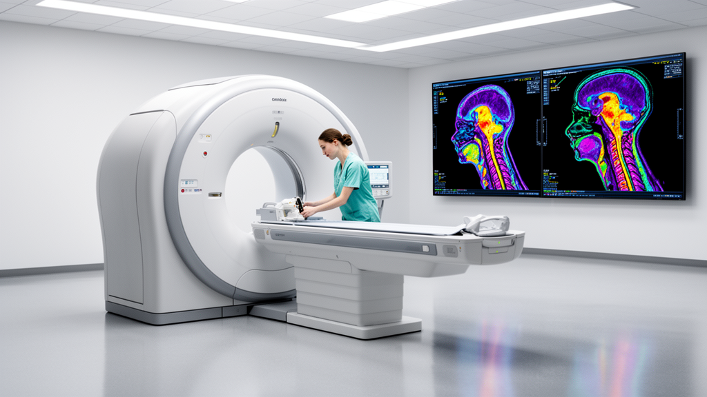
A retrospective clinical study reveals that routine adherence to standardized quality control procedures serves as the primary operational factor determining the diagnostic performance and radiation dosing consistency of SPECT/CT in detecting ectopic parathyroid adenomas. Investigating a cohort of 136 adult patients presenting with suspected primary hyperparathyroidism at Al-Hada Armed Forces Hospital, researchers evaluated how technical discipline and radiopharmaceutical selection interact during dual-phase scintigraphy. The investigation demonstrated that rigorous daily quality control produced a 97.1 percent lesion detection rate across examined cases, whereas operational lapses and undocumented calibration reduced overall diagnostic accuracy to 72.1 percent. Although both technetium-99m sestamibi and technetium-99m tetrofosmin delivered reliable diagnostic yield under strictly monitored imaging conditions, sestamibi demonstrated superior resilience whenever quality control standards slipped, preserving acceptable detection where alternative tracer performance degraded sharply.
Preoperative localization of diseased parathyroid tissue represents a critical clinical objective in managing primary hyperparathyroidism, where precise surgical excision directly depends on accurate non-invasive imaging. Hybrid single-photon emission computed tomography integrated with low-dose non-contrast computed tomography has become the standard modality for parathyroid mapping because it combines functional metabolic information with high-resolution cross-sectional anatomical detail. While emerging positron emission tomography techniques such as fluorine-18 fluorocholine offer notable capabilities in challenging diagnostic scenarios, widespread adoption remains constrained by substantial expense, restricted availability, and limited institutional access. Consequently, hybrid SPECT/CT continues to serve as the most practical primary imaging tool for surgical guidance. Nevertheless, clinical outcomes in nuclear medicine departments vary depending on operational rigor, camera calibration protocols, dose calibrator stability, and the specific pharmacokinetic properties of administered radiopharmaceutical agents utilized during dual-phase examinations.
The radiopharmaceuticals routinely utilized in parathyroid scintigraphy comprise technetium-99m sestamibi, designated MIBI, and technetium-99m tetrofosmin, commercialized as Myoview. Despite sharing this uptake mechanism, the two radiotracers display distinct pharmacokinetic clearance properties that significantly influence image acquisition timing and target-to-background contrast. Sestamibi features pronounced intracellular mitochondrial retention accompanied by relatively slow tissue washout, which sustains durable target contrast throughout protracted dual-phase scanning protocols. In contrast, tetrofosmin exhibits accelerated clearance and rapid tissue transit, an attribute that shortens examination intervals but simultaneously heightens vulnerability to procedural timing discrepancies and premature washout. These physiological differences render tetrofosmin particularly sensitive to operational variations, equipment misalignments, or protocol inconsistencies during delayed diagnostic imaging sessions.
Crucially, the investigative cohort was restricted entirely to patients presenting with surgically verified ectopic parathyroid adenomas, defined as abnormal hyperfunctioning glandular tissue positioned outside typical posterior thyroid anatomical spaces. These ectopic sites encompassed mediastinal, retroesophageal, intrathymic, and carotid sheath locations, representing challenging presentations where conventional planar imaging frequently falters. Because cases involving classic orthotopic posterior thyroid locations were intentionally excluded from analysis, every diagnostic detection and localization metric reported specifically reflects performance against surgically confirmed ectopic pathology. Operative exploration findings alongside formal histopathological verification served as the diagnostic reference standard against which all pre-surgical scintigraphic interpretations and hybrid SPECT/CT localization coordinates were systematically validated.
The clinical cohort of 136 individuals was evenly divided into four discrete analytical groups of 34 patients each, structured around the specific radiopharmaceutical administered and institutional quality control compliance. Group one received technetium-99m sestamibi under regular quality control, group two received technetium-99m tetrofosmin under regular quality control, group three received sestamibi under irregular quality control, and group four received tetrofosmin under irregular quality control. The standardized imaging workflow encompassed dual-phase image acquisition, commencing with early planar acquisitions obtained between 15 and 30 minutes following intravenous tracer injection, followed by delayed planar acquisitions scheduled at 90 to 150 minutes. Immediately following delayed planar imaging, patients underwent hybrid SPECT/CT scanning on a dual-head gamma camera equipped with low-dose non-contrast computed tomography for attenuation correction and structural registration. Regular quality control demanded verified daily gamma camera uniformity checks, dose calibrator constancy testing, and compliance with standardized acquisition settings.
Assessment of administered radiopharmaceutical activities revealed that rigorous quality control practices fostered tighter dosing precision and reproducible activity delivery across patient groups. Patients in group one administered sestamibi under regular quality control received the highest mean injected activity at 748.06 megabecquerels, accompanied by a standard deviation of 13.60 megabecquerels. Group three, imaged with sestamibi under irregular quality control conditions, recorded a mean administered activity of 712.62 megabecquerels with a standard deviation of 11.07 megabecquerels. For tetrofosmin, group two under regular quality control registered an average activity of 709.85 megabecquerels with a narrow standard deviation of 9.49 megabecquerels, whereas group four under irregular quality control averaged 692.65 megabecquerels with a standard deviation of 13.20 megabecquerels. Although sestamibi exhibited a marginally broader standard deviation under regular oversight due to wider institutional prescribing ranges, regular quality control demonstrated superior overall dose confinement across both compounds.
Detailed stratification of administered radiopharmaceutical dosages across patient demographic subsets confirmed that routine quality control procedures maintain equitable activity delivery irrespective of sex or chronological age. Under regular quality control environments, male patients within group one received an average of 750.40 megabecquerels while female counterparts received 746.21 megabecquerels; similarly, group two males received 708.75 megabecquerels compared with 710.83 megabecquerels in females. In contrast, irregular quality control groups exhibited expanded variations, exemplified by group four where male patients received 695.33 megabecquerels while females received 690.53 megabecquerels, although sex-based differences remained minor and lacked statistical significance. Age-stratified analyses comparing patients under 55 years against those aged 55 and older demonstrated comparable dosing stability under disciplined oversight, recording 745.27 versus 749.39 megabecquerels in group one and 706.09 versus 711.65 megabecquerels in group two. Such standardized dosing uniformity provides essential baseline data required to establish reliable local Diagnostic Reference Levels.
Diagnostic performance metrics confirmed that operational quality control adherence exerts a profound and statistically significant influence on the primary detection of ectopic parathyroid adenomas. When analyzing the entire cohort across all tracer types, SPECT/CT examinations conducted under regular quality control conditions achieved an overall lesion detection rate of 97.1 percent, identifying 66 out of 68 confirmed lesions. Conversely, under irregular quality control conditions where instrument calibration and protocol documentation lapsed, collective detection plummeted to 72.1 percent, representing only 49 successful identifications out of 68 cases. Statistical evaluation confirmed this discrepancy was highly significant, yielding a chi-square statistic of 16.275 with a p-value below 0.001. The investigators emphasized that image degradation stemming from energy peaking drift, uncorrected field non-uniformities, and unstandardized reconstruction filters directly eroded target-to-background contrast, precipitating diagnostic uncertainty and causing clinicians to miss abnormal parathyroid tissue that was readily visible under calibrated instrument conditions.
The deleterious consequences of unstandardized operational practices proved even more pronounced when evaluating precise anatomical localization accuracy rather than mere binary adenoma detection. Correct localization demanded that the hybrid imaging examination accurately map the ectopic glandular lesion to its exact anatomical compartment as subsequently verified during surgical intervention. Across regular quality control cohorts, SPECT/CT successfully localized 79 percent of all confirmed ectopic lesions, providing surgeons with dependable coordinates for targeted operative approaches. Under irregular quality control, however, correct anatomical localization collapsed dramatically to a mere 21 percent, meaning nearly four out of five lesions suffered spatial mischaracterization. Individual subgroup analysis showed that group one with sestamibi under regular quality control achieved the highest localization rate at 88.2 percent, followed by group two with tetrofosmin under regular oversight at 70.6 percent. In sharp contrast, irregular quality control reduced localization accuracy to 29.4 percent in group three and 23.5 percent in group four.
Comparing radiopharmaceutical agents under identical operational constraints revealed a distinct divergence in tracer resilience whenever quality control protocols were compromised. Under regular quality control, both imaging agents performed admirably, with sestamibi attaining a perfect 100 percent detection rate in group one and tetrofosmin achieving a robust 94.1 percent detection rate in group two. However, when quality control discipline lapsed, tetrofosmin experienced catastrophic performance degradation, dropping to a 58.8 percent detection rate alongside a 76.5 percent localization error rate in group four. Sestamibi exhibited markedly superior stability in group three, sustaining an 85.3 percent detection rate despite incurring a 70.6 percent localization error rate under degraded imaging environments. This disparity in diagnostic durability aligns with the established mitochondrial retention kinetics of sestamibi, which maintains sufficient parathyroid tissue activity to compensate for minor technical imperfections, whereas tetrofosmin’s rapid clearance renders it acutely vulnerable to misregistration and contrast loss during procedural drift.
Throughout the 136-patient investigation, hybrid SPECT/CT proved overwhelmingly superior to conventional planar scintigraphy across all diagnostic parameters and patient cohorts. Overall, SPECT/CT achieved an aggregate detection rate of 84.6 percent, accurately identifying 115 ectopic adenomas, whereas planar scintigraphy detected only 36.0 percent, or 49 cases. Furthermore, planar imaging performance remained completely insensitive to radiopharmaceutical choice, yielding statistically indistinguishable detection rates of 39.7 percent for sestamibi and 32.4 percent for tetrofosmin with a chi-square statistic of 0.798 and a p-value of 0.372. Planar scintigraphy completely lacked the spatial resolution, depth recovery, and computed tomography attenuation correction necessary to resolve deeply seated mediastinal or retroesophageal ectopic adenomas. The study authors concluded that nuclear medicine departments must implement strict quality control standards, including daily camera uniformity checks and locked reconstruction settings, while prioritizing sestamibi whenever technical variability cannot be eliminated, thereby maximizing surgical success in parathyroid localization.
Subject of Research: Impact of quality control practices and radiopharmaceutical choice on parathyroid SPECT/CT diagnostic performance and administered dose consistency
Article Title: Diagnostic accuracy of SPECT/CT in parathyroid adenoma detection: impact of radiopharmaceutical choice and quality control practices
Article References: Alasmari, B., Sherif, R. S. E.-D., Ghannam, M. M., Elsaban, K., & Al-Amri, M. A. (2026). Diagnostic accuracy of SPECT/CT in parathyroid adenoma detection: impact of radiopharmaceutical choice and quality control practices. EJNMMI Reports, 10(1), Article 12. https://doi.org/10.1186/s41824-026-00291-x
Image Credits: AI Generated
DOI: 10.1186/s41824-026-00291-x
Keywords: SPECT/CT, parathyroid adenoma, quality control, technetium-99m sestamibi, technetium-99m tetrofosmin, dual-phase scintigraphy, planar scintigraphy, diagnostic accuracy, administered activity, ectopic parathyroid, primary hyperparathyroidism, diagnostic reference levels, gamma camera uniformity, dose calibrator constancy, radiation dose consistency, surgical localization
News Source: Ophelia Keating. (November 13, 2026). Rigorous Quality Control Dictates SPECT/CT Accuracy in Parathyroid Adenoma Localization. Scienmag.